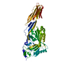
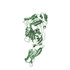
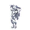
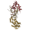
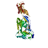
- PDB-3hvn: Crystal structure of cytotoxin protein suilysin from Streptococcu... -
+
Open data
ID or keywords:
Loading...
-
Basic information
Entry
Database: PDB / ID: 3hvn
Title
Crystal structure of cytotoxin protein suilysin from Streptococcus suis
Components
Hemolysin
Keywords
TOXIN / beta-strand rich / elongated rod like / pore forming
Function / homology
Function and homology information
: / hemolysis in another organism / cholesterol binding / membrane => GO:0016020 / toxin activity / killing of cells of another organism / host cell plasma membrane / extracellular region Similarity search - Function
Redundancy: 9.5 % / Av σ(I) over netI: 30.36 / Number: 122778 / Rmerge(I) obs: 0.082 / Χ2: 2.33 / D res high: 3 Å / D res low: 50 Å / Num. obs: 12949 / % possible obs: 98.2
Diffraction reflection shell
Highest resolution (Å)
Lowest resolution (Å)
% possible obs (%)
ID
Rmerge(I) obs
Chi squared
Redundancy
6.46
50
97
1
0.068
7.972
7.7
5.13
6.46
100
1
0.071
4.112
9.9
4.48
5.13
99.7
1
0.07
2.999
10.3
4.07
4.48
99.9
1
0.072
2.076
10.5
3.78
4.07
100
1
0.094
1.51
10.8
3.56
3.78
100
1
0.126
1.445
9.8
3.38
3.56
100
1
0.161
1.036
10.9
3.23
3.38
100
1
0.221
0.883
10.1
3.11
3.23
98.7
1
0.3
0.743
8.3
3
3.11
86.6
1
0.322
0.774
6.2
Reflection
Resolution: 2.85→50 Å / Num. obs: 13204 / % possible obs: 88.5 % / Redundancy: 5.7 % / Rmerge(I) obs: 0.052 / Χ2: 1.033 / Net I/σ(I): 30.364
Reflection shell
Resolution (Å)
Redundancy (%)
Rmerge(I) obs
Num. unique all
Χ2
% possible all
2.85-2.95
5.6
0.316
968
0.864
66.4
2.95-3.07
5.8
0.233
1234
0.926
83.7
3.07-3.21
5.8
0.171
1318
1.026
90.8
3.21-3.38
5.7
0.107
1396
1.186
94.3
3.38-3.59
5.8
0.075
1375
1.15
93.9
3.59-3.87
5.7
0.063
1400
1.021
93.8
3.87-4.26
5.7
0.052
1377
0.985
93.5
4.26-4.87
5.8
0.044
1400
1.109
92.4
4.87-6.14
5.8
0.041
1358
1.062
90.2
6.14-50
5.6
0.043
1378
0.931
85.5
-
Phasing
Phasing
Method: MAD
-
Processing
Software
Name
Version
Classification
NB
DENZO
datareduction
SCALEPACK
datascaling
SOLVE
phasing
RESOLVE
phasing
PHENIX
refinement
PDB_EXTRACT
3.005
dataextraction
Refinement
Method to determine structure: MAD / Resolution: 2.852→28.409 Å / Occupancy max: 1 / Occupancy min: 1 / SU ML: 0.74 / σ(F): 0.16 / Stereochemistry target values: MLHL
Rfactor
Num. reflection
% reflection
Rfree
0.294
637
5.03 %
Rwork
0.279
-
-
obs
0.28
12653
85.07 %
Solvent computation
Shrinkage radii: 0.9 Å / VDW probe radii: 1.11 Å / Solvent model: FLAT BULK SOLVENT MODEL / Bsol: 91.358 Å2 / ksol: 0.298 e/Å3
In the structure databanks used in Yorodumi, some data are registered as the other names, "COVID-19 virus" and "2019-nCoV". Here are the details of the virus and the list of structure data.
Jan 31, 2019. EMDB accession codes are about to change! (news from PDBe EMDB page)
EMDB accession codes are about to change! (news from PDBe EMDB page)
The allocation of 4 digits for EMDB accession codes will soon come to an end. Whilst these codes will remain in use, new EMDB accession codes will include an additional digit and will expand incrementally as the available range of codes is exhausted. The current 4-digit format prefixed with “EMD-” (i.e. EMD-XXXX) will advance to a 5-digit format (i.e. EMD-XXXXX), and so on. It is currently estimated that the 4-digit codes will be depleted around Spring 2019, at which point the 5-digit format will come into force.
The EM Navigator/Yorodumi systems omit the EMD- prefix.
Related info.:Q: What is EMD? / ID/Accession-code notation in Yorodumi/EM Navigator
Yorodumi is a browser for structure data from EMDB, PDB, SASBDB, etc.
This page is also the successor to EM Navigator detail page, and also detail information page/front-end page for Omokage search.
The word "yorodu" (or yorozu) is an old Japanese word meaning "ten thousand". "mi" (miru) is to see.
Related info.:EMDB / PDB / SASBDB / Comparison of 3 databanks / Yorodumi Search / Aug 31, 2016. New EM Navigator & Yorodumi / Yorodumi Papers / Jmol/JSmol / Function and homology information / Changes in new EM Navigator and Yorodumi
 Movie
Movie Controller

 Yorodumi
 Open data
 Basic information
 Components
 Keywords
 Function and homology information
 Streptococcus suis (bacteria)
 X-RAY DIFFRACTION /
  Authors
 Citation
 Structure visualization
 Downloads & links
 Other downloads
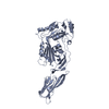


 PDBj
PDBj
 Assembly
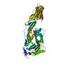
 Mass: 168.038 Da / Num. of mol.: 1 / Source method: obtained synthetically / Formula: C3H2F6O
 Mass: 148.200 Da / Num. of mol.: 1 / Source method: obtained synthetically / Formula: C7H16O3
 Mass: 18.015 Da / Num. of mol.: 7 / Source method: isolated from a natural source / Formula: H2O
 Sample preparation
 Processing