ムービー
ムービー コントローラー


+ データを開く

- 基本情報
基本情報
| 登録情報 | データベース: EMDB / ID: EMD-3849 | |||||||||
|---|---|---|---|---|---|---|---|---|---|---|
| タイトル | Single particle cryo-STEM of ferritin with Fe at low stoichiometry | |||||||||
 マップデータ マップデータ | Ferritin with 100 Fe atoms from single particle cryo-STEM data | |||||||||
 試料 試料 |
| |||||||||
| 生物種 |  Homo sapiens (ヒト) Homo sapiens (ヒト) | |||||||||
| 手法 | 単粒子再構成法 / クライオ電子顕微鏡法 / 解像度: 21.0 Å | |||||||||
 データ登録者 データ登録者 | Elad N / Bellapadrona G / Houben L / Sagi I / Elbaum M | |||||||||
 引用 引用 | ジャーナル: Proc Natl Acad Sci U S A / 年: 2017 タイトル: Detection of isolated protein-bound metal ions by single-particle cryo-STEM. 著者: Nadav Elad / Giuliano Bellapadrona / Lothar Houben / Irit Sagi / Michael Elbaum /  要旨: Metal ions play essential roles in many aspects of biological chemistry. Detecting their presence and location in proteins and cells is important for understanding biological function. Conventional ...Metal ions play essential roles in many aspects of biological chemistry. Detecting their presence and location in proteins and cells is important for understanding biological function. Conventional structural methods such as X-ray crystallography and cryo-transmission electron microscopy can identify metal atoms on protein only if the protein structure is solved to atomic resolution. We demonstrate here the detection of isolated atoms of Zn and Fe on ferritin, using cryogenic annular dark-field scanning transmission electron microscopy (cryo-STEM) coupled with single-particle 3D reconstructions. Zn atoms are found in a pattern that matches precisely their location at the ferroxidase sites determined earlier by X-ray crystallography. By contrast, the Fe distribution is smeared along an arc corresponding to the proposed path from the ferroxidase sites to the mineral nucleation sites along the twofold axes. In this case the single-particle reconstruction is interpreted as a probability distribution function based on the average of individual locations. These results establish conditions for detection of isolated metal atoms in the broader context of electron cryo-microscopy and tomography. | |||||||||
| 履歴 |
|
- 構造の表示
構造の表示
| ムービー |
 ムービービューア ムービービューア |
|---|---|
| 構造ビューア | EMマップ: SurfViewMolmilJmol/JSmol |
| 添付画像 |

- ダウンロードとリンク
-EMDBアーカイブ
| マップデータ | emd_3849.map.gz | 925 KB | EMDBマップデータ形式 | |
|---|---|---|---|---|
| ヘッダ (付随情報) | emd-3849-v30.xmlemd-3849.xml | 12.2 KB 12.2 KB | 表示 表示 | EMDBヘッダ |
| 画像 | 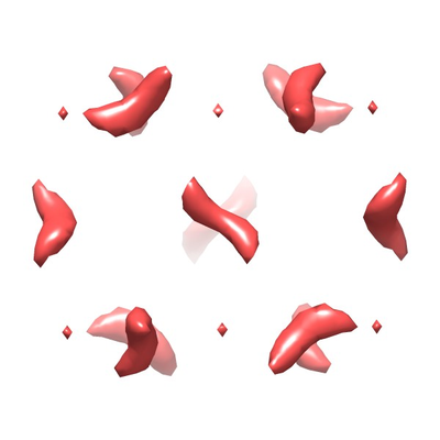 emd_3849.png emd_3849.png | 76.8 KB | ||
| アーカイブディレクトリ |  http://ftp.pdbj.org/pub/emdb/structures/EMD-3849ftp://ftp.pdbj.org/pub/emdb/structures/EMD-3849 http://ftp.pdbj.org/pub/emdb/structures/EMD-3849ftp://ftp.pdbj.org/pub/emdb/structures/EMD-3849 | HTTPS FTP |
-検証レポート
| 文書・要旨 | emd_3849_validation.pdf.gz | 216.8 KB | 表示 | EMDB検証レポート |
|---|---|---|---|---|
| 文書・詳細版 | emd_3849_full_validation.pdf.gz | 215.9 KB | 表示 | |
| XML形式データ | emd_3849_validation.xml.gz | 4.3 KB | 表示 | |
| アーカイブディレクトリ | https://ftp.pdbj.org/pub/emdb/validation_reports/EMD-3849ftp://ftp.pdbj.org/pub/emdb/validation_reports/EMD-3849 | HTTPS FTP |
-関連構造データ
-リンク
| EMDBのページ | EMDB (EBI/PDBe) / EMDataResource |
|---|
-マップ
| ファイル | ダウンロード / ファイル: emd_3849.map.gz / 形式: CCP4 / 大きさ: 2 MB / タイプ: IMAGE STORED AS FLOATING POINT NUMBER (4 BYTES) | ||||||||||||||||||||||||||||||||||||||||||||||||||||||||||||
|---|---|---|---|---|---|---|---|---|---|---|---|---|---|---|---|---|---|---|---|---|---|---|---|---|---|---|---|---|---|---|---|---|---|---|---|---|---|---|---|---|---|---|---|---|---|---|---|---|---|---|---|---|---|---|---|---|---|---|---|---|---|
| 注釈 | Ferritin with 100 Fe atoms from single particle cryo-STEM data | ||||||||||||||||||||||||||||||||||||||||||||||||||||||||||||
| 投影像・断面図 | 画像のコントロール
画像は Spider により作成 | ||||||||||||||||||||||||||||||||||||||||||||||||||||||||||||
| ボクセルのサイズ | X=Y=Z: 3.3 Å | ||||||||||||||||||||||||||||||||||||||||||||||||||||||||||||
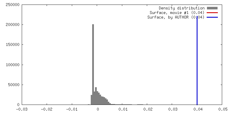
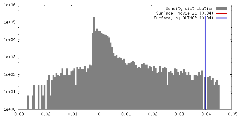
| 密度 |
| ||||||||||||||||||||||||||||||||||||||||||||||||||||||||||||
| 対称性 | 空間群: 1 | ||||||||||||||||||||||||||||||||||||||||||||||||||||||||||||
| 詳細 | EMDB XML:
CCP4マップ ヘッダ情報:
| ||||||||||||||||||||||||||||||||||||||||||||||||||||||||||||
 Z (Sec.)
 Y (Row.)
 X (Col.)


-添付データ
- 試料の構成要素
-全体 : Human heavy chain ferritin with Fe at low stoichiometry
| 全体 | 名称: Human heavy chain ferritin with Fe at low stoichiometry |
|---|---|
| 要素 |
|
-超分子 #1: Human heavy chain ferritin with Fe at low stoichiometry
| 超分子 | 名称: Human heavy chain ferritin with Fe at low stoichiometry タイプ: complex / ID: 1 / 親要素: 0 詳細: Bacterially expressed and purified human heavy chain ferritin mixed with a 100 fold molar excess of Fe(II)SO4 |
|---|---|
| 由来(天然) | 生物種: Homo sapiens (ヒト) |
| 組換発現 | 生物種:  |
| 分子量 | 理論値: 504 KDa |
-実験情報
-構造解析
| 手法 | クライオ電子顕微鏡法 |
|---|---|
 解析 解析 | 単粒子再構成法 |
| 試料の集合状態 | particle |
-試料調製
| 濃度 | 0.13 mg/mL | ||||||
|---|---|---|---|---|---|---|---|
| 緩衝液 | pH: 7.2 / 構成要素:
| ||||||
| グリッド | モデル: Quantifoil R2/1 / 材質: COPPER / メッシュ: 200 / 支持フィルム - #0 - Film type ID: 1 / 支持フィルム - #0 - 材質: CARBON / 支持フィルム - #0 - トポロジー: HOLEY / 支持フィルム - #1 - Film type ID: 2 / 支持フィルム - #1 - 材質: CARBON / 支持フィルム - #1 - トポロジー: CONTINUOUS / 前処理 - タイプ: GLOW DISCHARGE / 前処理 - 雰囲気: AIR | ||||||
| 凍結 | 凍結剤: ETHANE / チャンバー内湿度: 95 % / チャンバー内温度: 297 K / 装置: LEICA EM GP | ||||||
| 詳細 | For metal loading, stock protein (2.9 mg/ml) was diluted to 0.25 mg/ml and mixed with a 100 fold molar excess of Fe(II)SO4. |
- 電子顕微鏡法
| 顕微鏡 | FEI TECNAI F20 |
|---|---|
| 詳細 | STEM mode imaging using Fischione model 3000 HAADF detector. Camera length: 520 mm. |
| 撮影 | フィルム・検出器のモデル: OTHER / デジタル化 - サイズ - 横: 2048 pixel / デジタル化 - サイズ - 縦: 2048 pixel / 撮影したグリッド数: 1 / 実像数: 58 / 平均電子線量: 127.0 e/Å2 詳細: ADF images collected with a Fischione Model 3000 high-angle annular DF detector |
| 電子線 | 加速電圧: 200 kV / 電子線源:  FIELD EMISSION GUN FIELD EMISSION GUN |
| 電子光学系 | C2レンズ絞り径: 70.0 µm / 照射モード: OTHER / 撮影モード: OTHER / Cs: 2.0 mm / 倍率(公称値): 450000 |
| 試料ステージ | 試料ホルダーモデル: GATAN 626 SINGLE TILT LIQUID NITROGEN CRYO TRANSFER HOLDER ホルダー冷却材: NITROGEN |
| 実験機器 |  モデル: Tecnai F20 / 画像提供: FEI Company |