 Movie
Movie Controller


+ Open data

- Basic information
| Entry |  | |||||||||
|---|---|---|---|---|---|---|---|---|---|---|
| Title | Dimeric tail tube protein gpVs of bacteriophage lambda | |||||||||
 Map data Map data | ||||||||||
 Sample Sample |
| |||||||||
 Keywords Keywords | Bacteriophage / caudovirales / siphoviridae / tail complex / delivery device / phage lambda / cryo-EM / VIRAL PROTEIN | |||||||||
| Function / homology |  Function and homology information Function and homology informationvirus tail, tube / symbiont genome ejection through host cell envelope, long flexible tail mechanism / viral tail assembly / host cell cytoplasm Similarity search - Function | |||||||||
| Biological species |  Escherichia phage Lambda (virus) / Escherichia phage lambda (virus) Escherichia phage Lambda (virus) / Escherichia phage lambda (virus) | |||||||||
| Method | single particle reconstruction / cryo EM / Resolution: 3.8 Å | |||||||||
 Authors Authors | Wang JW | |||||||||
| Funding support |  China, 1 items China, 1 items
| |||||||||
 Citation Citation | Journal: Structure / Year: 2024 Title: Architecture of the bacteriophage lambda tail. Authors: Chang Wang / Jinsong Duan / Zhiwei Gu / Xiaofei Ge / Jianwei Zeng / Jiawei Wang / Abstract: Bacteriophage lambda has a double-stranded DNA genome and a long, flexible, non-contractile tail encoded by a contiguous block of 11 genes downstream of the head genes. The tail allows host ...Bacteriophage lambda has a double-stranded DNA genome and a long, flexible, non-contractile tail encoded by a contiguous block of 11 genes downstream of the head genes. The tail allows host recognition and delivery of viral DNA from the head shell to the cytoplasm of the infected cell. Here, we present a high-resolution structure of the tail complex of bacteriophage lambda determined by cryoelectron microscopy. Most component proteins of the lambda tail were determined at the atomic scale. The structure sheds light on the molecular organization of the extensively studied tail of bacteriophage lambda. | |||||||||
| History |
|
- Structure visualization
| Supplemental images |
|---|

- Downloads & links
-EMDB archive
| Map data | emd_37218.map.gz | 59.3 MB | EMDB map data format | |
|---|---|---|---|---|
| Header (meta data) | emd-37218-v30.xmlemd-37218.xml | 13.6 KB 13.6 KB | Display Display | EMDB header |
| FSC (resolution estimation) | emd_37218_fsc.xml | 8.5 KB | Display | FSC data file |
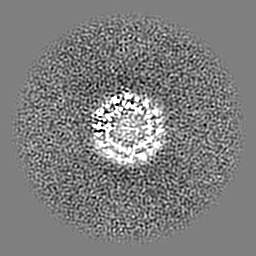
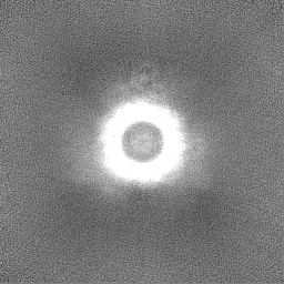
| Images | 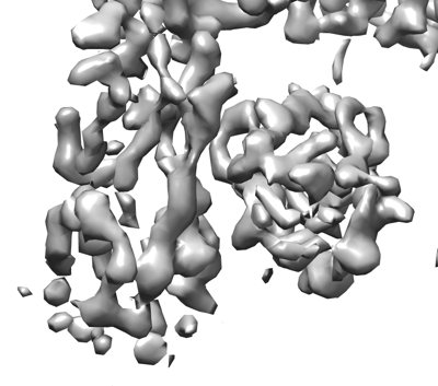 emd_37218.png emd_37218.png | 88.2 KB | ||
| Filedesc metadata | emd-37218.cif.gz | 4.9 KB | ||
| Others | emd_37218_half_map_1.map.gzemd_37218_half_map_2.map.gz | 59.3 MB 59.3 MB | ||
| Archive directory |  http://ftp.pdbj.org/pub/emdb/structures/EMD-37218ftp://ftp.pdbj.org/pub/emdb/structures/EMD-37218 http://ftp.pdbj.org/pub/emdb/structures/EMD-37218ftp://ftp.pdbj.org/pub/emdb/structures/EMD-37218 | HTTPS FTP |
-Validation report
| Summary document | emd_37218_validation.pdf.gz | 1.2 MB | Display | EMDB validaton report |
|---|---|---|---|---|
| Full document | emd_37218_full_validation.pdf.gz | 1.2 MB | Display | |
| Data in XML | emd_37218_validation.xml.gz | 16.1 KB | Display | |
| Data in CIF | emd_37218_validation.cif.gz | 20.5 KB | Display | |
| Arichive directory | https://ftp.pdbj.org/pub/emdb/validation_reports/EMD-37218ftp://ftp.pdbj.org/pub/emdb/validation_reports/EMD-37218 | HTTPS FTP |
-Related structure data
| Related structure data | 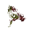 8kgeMC 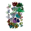 8iydC  8iykC  8iylC  8jvmC C: citing same article ( M: atomic model generated by this map |
|---|---|
| Similar structure data |
-Links
| EMDB pages | EMDB (EBI/PDBe) / EMDataResource |
|---|
-Map
| File | Download / File: emd_37218.map.gz / Format: CCP4 / Size: 64 MB / Type: IMAGE STORED AS FLOATING POINT NUMBER (4 BYTES) | ||||||||||||||||||||||||||||||||||||
|---|---|---|---|---|---|---|---|---|---|---|---|---|---|---|---|---|---|---|---|---|---|---|---|---|---|---|---|---|---|---|---|---|---|---|---|---|---|
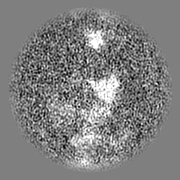
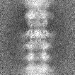
| Projections & slices | Image control
Images are generated by Spider. | ||||||||||||||||||||||||||||||||||||
| Voxel size | X=Y=Z: 1.0742 Å | ||||||||||||||||||||||||||||||||||||
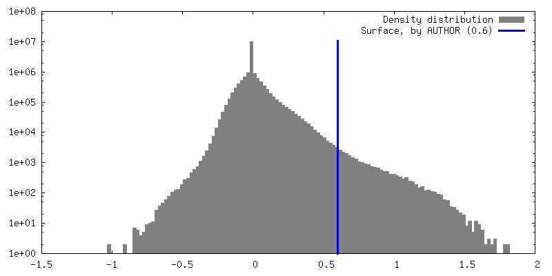
| Density |
| ||||||||||||||||||||||||||||||||||||
| Symmetry | Space group: 1 | ||||||||||||||||||||||||||||||||||||
| Details | EMDB XML:
|
 Z (Sec.)
 Y (Row.)
 X (Col.)


-Supplemental data
-Half map: #1
| File | emd_37218_half_map_1.map | ||||||||||||
|---|---|---|---|---|---|---|---|---|---|---|---|---|---|
| Projections & Slices |
| ||||||||||||
| Density Histograms |


-Half map: #2
| File | emd_37218_half_map_2.map | ||||||||||||
|---|---|---|---|---|---|---|---|---|---|---|---|---|---|
| Projections & Slices |
| ||||||||||||
| Density Histograms |


- Sample components
-Entire : Bacteriophage lambda tail
| Entire | Name: Bacteriophage lambda tail |
|---|---|
| Components |
|
-Supramolecule #1: Bacteriophage lambda tail
| Supramolecule | Name: Bacteriophage lambda tail / type: complex / ID: 1 / Parent: 0 / Macromolecule list: all |
|---|---|
| Source (natural) | Organism: Escherichia phage Lambda (virus) |
-Macromolecule #1: Tail tube protein
| Macromolecule | Name: Tail tube protein / type: protein_or_peptide / ID: 1 / Number of copies: 2 / Enantiomer: LEVO |
|---|---|
| Source (natural) | Organism: Escherichia phage lambda (virus) |
| Molecular weight | Theoretical: 25.831779 KDa |
| Recombinant expression | Organism:  |
| Sequence | String: MPVPNPTMPV KGAGTTLWVY KGSGDPYANP LSDVDWSRLA KVKDLTPGEL TAESYDDSYL DDEDADWTAT GQGQKSAGDT SFTLAWMPG EQGQQALLAW FNEGDTRAYK IRFPNGTVDV FRGWVSSIGK AVTAKEVITR TVKVTNVGRP SMAEDRSTVT A ATGMTVTP ...String: MPVPNPTMPV KGAGTTLWVY KGSGDPYANP LSDVDWSRLA KVKDLTPGEL TAESYDDSYL DDEDADWTAT GQGQKSAGDT SFTLAWMPG EQGQQALLAW FNEGDTRAYK IRFPNGTVDV FRGWVSSIGK AVTAKEVITR TVKVTNVGRP SMAEDRSTVT A ATGMTVTP ASTSVVKGQS TTLTVAFQPE GVTDKSFRAV SADKTKATVS VSGMTITVNG VAAGKVNIPV VSGNGEFAAV AE ITVTAS UniProtKB: Tail tube protein |
-Experimental details
-Structure determination
| Method | cryo EM |
|---|---|
 Processing Processing | single particle reconstruction |
| Aggregation state | particle |
-Sample preparation
| Buffer | pH: 7 |
|---|---|
| Vitrification | Cryogen name: NITROGEN |
- Electron microscopy
| Microscope | FEI TITAN KRIOS |
|---|---|
| Image recording | Film or detector model: GATAN K3 (6k x 4k) / Average electron dose: 50.0 e/Å2 |
| Electron beam | Acceleration voltage: 300 kV / Electron source:  FIELD EMISSION GUN FIELD EMISSION GUN |
| Electron optics | Illumination mode: FLOOD BEAM / Imaging mode: BRIGHT FIELD / Nominal defocus max: 2.0 µm / Nominal defocus min: 1.0 µm |
| Experimental equipment |  Model: Titan Krios / Image courtesy: FEI Company |

