 Movie
Movie Controller


+ Open data

- Basic information
| Entry |  | |||||||||
|---|---|---|---|---|---|---|---|---|---|---|
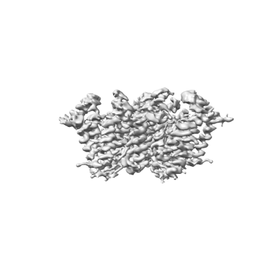
| Title | Membrane bound PRTase, C3 symmetry, acceptor bound | |||||||||
 Map data Map data | EM map of a membrane bound PRTase, C3 symmetry, acceptor bound | |||||||||
 Sample Sample |
| |||||||||
 Keywords Keywords | phosphoribose transferase complex / MEMBRANE PROTEIN | |||||||||
| Function / homology |  Function and homology information Function and homology informationdecaprenyl-phosphate phosphoribosyltransferase / arabinosyltransferase activity / glycolipid biosynthetic process / capsule polysaccharide biosynthetic process / transferase activity, transferring alkyl or aryl (other than methyl) groups / cell wall organization / transferase activity / magnesium ion binding / plasma membrane Similarity search - Function | |||||||||
| Biological species |  Mycobacterium tuberculosis H37Rv (bacteria) / Mycobacterium tuberculosis (strain ATCC 25618 / H37Rv) (bacteria) Mycobacterium tuberculosis H37Rv (bacteria) / Mycobacterium tuberculosis (strain ATCC 25618 / H37Rv) (bacteria) | |||||||||
| Method | single particle reconstruction / cryo EM / Resolution: 3.36 Å | |||||||||
 Authors Authors | Wu FY / Gao S / Zhang L / Rao ZH | |||||||||
| Funding support |  China, 1 items China, 1 items
| |||||||||
 Citation Citation | Journal: Nat Microbiol / Year: 2024 Title: Structural analysis of phosphoribosyltransferase-mediated cell wall precursor synthesis in Mycobacterium tuberculosis. Authors: Shan Gao / Fangyu Wu / Sudagar S Gurcha / Sarah M Batt / Gurdyal S Besra / Zihe Rao / Lu Zhang /  Abstract: In Mycobacterium tuberculosis, Rv3806c is a membrane-bound phosphoribosyltransferase (PRTase) involved in cell wall precursor production. It catalyses pentosyl phosphate transfer from phosphoribosyl ...In Mycobacterium tuberculosis, Rv3806c is a membrane-bound phosphoribosyltransferase (PRTase) involved in cell wall precursor production. It catalyses pentosyl phosphate transfer from phosphoribosyl pyrophosphate to decaprenyl phosphate, to generate 5-phospho-β-ribosyl-1-phosphoryldecaprenol. Despite Rv3806c being an attractive drug target, structural and molecular mechanistic insight into this PRTase is lacking. Here we report cryogenic electron microscopy structures for Rv3806c in the donor- and acceptor-bound states. In a lipidic environment, Rv3806c is trimeric, creating a UbiA-like fold. Each protomer forms two helical bundles, which, alongside the bound lipids, are required for PRTase activity in vitro. Mutational and functional analyses reveal that decaprenyl phosphate and phosphoribosyl pyrophosphate bind the intramembrane and extramembrane cavities of Rv3806c, respectively, in a distinct manner to that of UbiA superfamily enzymes. Our data suggest a model for Rv3806c-catalysed phosphoribose transfer through an inverting mechanism. These findings provide a structural basis for cell wall precursor biosynthesis that could have potential for anti-tuberculosis drug development. | |||||||||
| History |
|
- Structure visualization
| Supplemental images |
|---|
- Downloads & links
-EMDB archive
| Map data | emd_36072.map.gz | 107 MB | EMDB map data format | |
|---|---|---|---|---|
| Header (meta data) | emd-36072-v30.xmlemd-36072.xml | 17.5 KB 17.5 KB | Display Display | EMDB header |
| Images |  emd_36072.png emd_36072.png | 41.7 KB | ||
| Filedesc metadata | emd-36072.cif.gz | 6.2 KB | ||
| Others | emd_36072_half_map_1.map.gzemd_36072_half_map_2.map.gz | 105.5 MB 105.5 MB | ||
| Archive directory |  http://ftp.pdbj.org/pub/emdb/structures/EMD-36072ftp://ftp.pdbj.org/pub/emdb/structures/EMD-36072 http://ftp.pdbj.org/pub/emdb/structures/EMD-36072ftp://ftp.pdbj.org/pub/emdb/structures/EMD-36072 | HTTPS FTP |
-Validation report
| Summary document | emd_36072_validation.pdf.gz | 831.9 KB | Display | EMDB validaton report |
|---|---|---|---|---|
| Full document | emd_36072_full_validation.pdf.gz | 831.5 KB | Display | |
| Data in XML | emd_36072_validation.xml.gz | 13.6 KB | Display | |
| Data in CIF | emd_36072_validation.cif.gz | 16 KB | Display | |
| Arichive directory | https://ftp.pdbj.org/pub/emdb/validation_reports/EMD-36072ftp://ftp.pdbj.org/pub/emdb/validation_reports/EMD-36072 | HTTPS FTP |
-Related structure data
| Related structure data |  8j8kMC 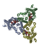 8j8jC M: atomic model generated by this map C: citing same article ( |
|---|---|
| Similar structure data |

-Links
| EMDB pages | EMDB (EBI/PDBe) / EMDataResource |
|---|
-Map
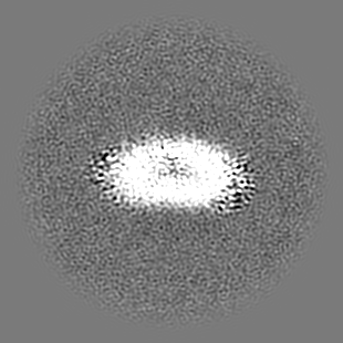
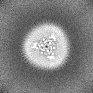
| File | Download / File: emd_36072.map.gz / Format: CCP4 / Size: 113.6 MB / Type: IMAGE STORED AS FLOATING POINT NUMBER (4 BYTES) | ||||||||||||||||||||||||||||||||||||
|---|---|---|---|---|---|---|---|---|---|---|---|---|---|---|---|---|---|---|---|---|---|---|---|---|---|---|---|---|---|---|---|---|---|---|---|---|---|
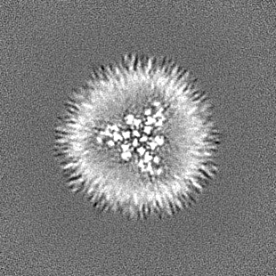
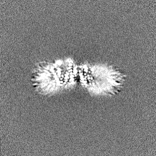
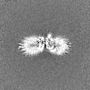
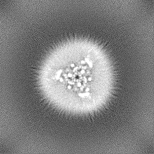
| Annotation | EM map of a membrane bound PRTase, C3 symmetry, acceptor bound | ||||||||||||||||||||||||||||||||||||
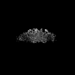
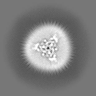
| Projections & slices | Image control
Images are generated by Spider. | ||||||||||||||||||||||||||||||||||||
| Voxel size | X=Y=Z: 0.96 Å | ||||||||||||||||||||||||||||||||||||
| Density |
| ||||||||||||||||||||||||||||||||||||
| Symmetry | Space group: 1 | ||||||||||||||||||||||||||||||||||||
| Details | EMDB XML:
|
 Z (Sec.)
 Y (Row.)
 X (Col.)


-Supplemental data
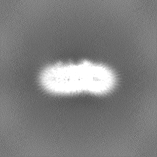
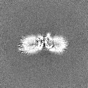
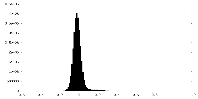
-Half map: EM half map A of a membrane bound PRTase, C3 symmetry, acceptor bound
| File | emd_36072_half_map_1.map | ||||||||||||
|---|---|---|---|---|---|---|---|---|---|---|---|---|---|
| Annotation | EM half map A of a membrane bound PRTase, C3 symmetry, acceptor bound | ||||||||||||
| Projections & Slices |
| ||||||||||||
| Density Histograms |
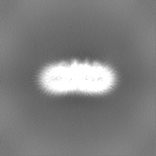


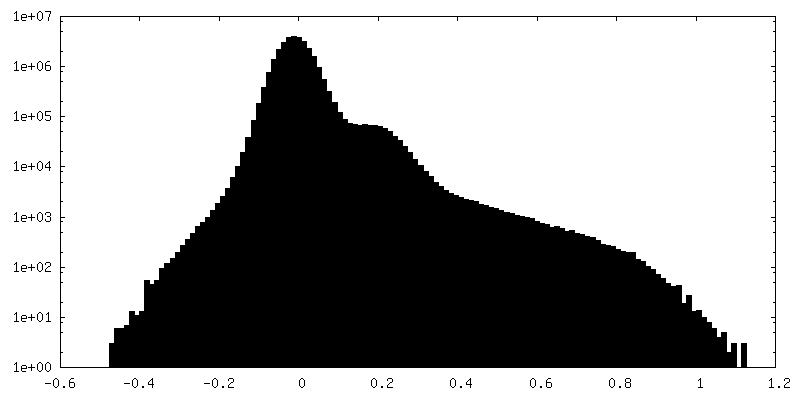
-Half map: EM half map B of a membrane bound PRTase, C3 symmetry, acceptor bound
| File | emd_36072_half_map_2.map | ||||||||||||
|---|---|---|---|---|---|---|---|---|---|---|---|---|---|
| Annotation | EM half map B of a membrane bound PRTase, C3 symmetry, acceptor bound | ||||||||||||
| Projections & Slices |
| ||||||||||||
| Density Histograms |
- Sample components
-Entire : trimeric phosphoribosyltransferase
| Entire | Name: trimeric phosphoribosyltransferase |
|---|---|
| Components |
|
-Supramolecule #1: trimeric phosphoribosyltransferase
| Supramolecule | Name: trimeric phosphoribosyltransferase / type: complex / ID: 1 / Parent: 0 / Macromolecule list: #1 Details: trimeric phosphoribosyltransferase, acceptor bound state |
|---|---|
| Source (natural) | Organism: Mycobacterium tuberculosis H37Rv (bacteria) |
| Molecular weight | Theoretical: 97.8 KDa |
-Macromolecule #1: Decaprenyl-phosphate phosphoribosyltransferase
| Macromolecule | Name: Decaprenyl-phosphate phosphoribosyltransferase / type: protein_or_peptide / ID: 1 / Number of copies: 3 / Enantiomer: LEVO / EC number: decaprenyl-phosphate phosphoribosyltransferase |
|---|---|
| Source (natural) | Organism: Mycobacterium tuberculosis (strain ATCC 25618 / H37Rv) (bacteria) |
| Molecular weight | Theoretical: 31.938473 KDa |
| Recombinant expression | Organism: Mycolicibacterium smegmatis MC2 155 (bacteria) |
| Sequence | String: MSEDVVTQPP ANLVAGVVKA IRPRQWVKNV LVLAAPLAAL GGGVRYDYVE VLSKVSMAFV VFSLAASAVY LVNDVRDVAA AAAAAAAAA AAAAAGVVPE WLAYTVAVVL GVTSLAGAWM LTPNLALVMV VYLAMQLAYC FGLKHQAVVE ICVVSSAYLI R AIAGGVAT ...String: MSEDVVTQPP ANLVAGVVKA IRPRQWVKNV LVLAAPLAAL GGGVRYDYVE VLSKVSMAFV VFSLAASAVY LVNDVRDVAA AAAAAAAAA AAAAAGVVPE WLAYTVAVVL GVTSLAGAWM LTPNLALVMV VYLAMQLAYC FGLKHQAVVE ICVVSSAYLI R AIAGGVAT KIPLSKWFLL IMAFGSLFMV AGKRYAELHL AERTGAAIRK SLESYTSTYL RFVWTLSATA VVLCYGLWAF ER DGYSGSW FAVSMIPFTI AILRYAVDVD GGLAGEPEDI ALRDRVLQLL ALAWIATVGA AVAFG UniProtKB: Decaprenyl-phosphate phosphoribosyltransferase |
-Macromolecule #2: MONO-TRANS, OCTA-CIS DECAPRENYL-PHOSPHATE
| Macromolecule | Name: MONO-TRANS, OCTA-CIS DECAPRENYL-PHOSPHATE / type: ligand / ID: 2 / Number of copies: 3 / Formula: DSL |
|---|---|
| Molecular weight | Theoretical: 779.165 Da |
| Chemical component information |  ChemComp-DSL: |
-Experimental details
-Structure determination
| Method | cryo EM |
|---|---|
 Processing Processing | single particle reconstruction |
| Aggregation state | particle |
-Sample preparation
| Concentration | 2.0 mg/mL | |||||||||
|---|---|---|---|---|---|---|---|---|---|---|
| Buffer | pH: 7.5 Component:
| |||||||||
| Grid | Model: Quantifoil R1.2/1.3 / Material: GOLD / Support film - Material: CARBON / Support film - topology: HOLEY / Pretreatment - Type: PLASMA CLEANING / Pretreatment - Time: 40 sec. | |||||||||
| Vitrification | Cryogen name: ETHANE / Chamber humidity: 100 % | |||||||||
| Details | this sample was mono disperse |
- Electron microscopy
| Microscope | FEI TITAN KRIOS |
|---|---|
| Specialist optics | Energy filter - Name: TFS Selectris |
| Image recording | Film or detector model: FEI FALCON IV (4k x 4k) / Number real images: 8894 / Average electron dose: 60.0 e/Å2 |
| Electron beam | Acceleration voltage: 300 kV / Electron source:  FIELD EMISSION GUN FIELD EMISSION GUN |
| Electron optics | Illumination mode: SPOT SCAN / Imaging mode: BRIGHT FIELD / Cs: 2.7 mm / Nominal defocus max: 2.0 µm / Nominal defocus min: 1.2 µm / Nominal magnification: 130000 |
| Sample stage | Specimen holder model: FEI TITAN KRIOS AUTOGRID HOLDER / Cooling holder cryogen: NITROGEN |
| Experimental equipment |  Model: Titan Krios / Image courtesy: FEI Company |
