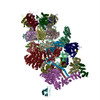
Movie
Movie Controller


+ Open data

- Basic information
| Entry | Database: EMDB / ID: EMD-3537 | ||||||||||||
|---|---|---|---|---|---|---|---|---|---|---|---|---|---|
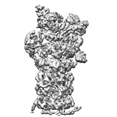
| Title | 26S proteasome in presence of BeFx (s4) | ||||||||||||
 Map data Map data | 26S proteasome in presence of BeFx (s4) | ||||||||||||
 Sample Sample |
| ||||||||||||
 Keywords Keywords | Macromolecular complex / 26S proteasome / Protease / Hydrolase | ||||||||||||
| Function / homology |  Function and homology information Function and homology informationnegative regulation of ERAD pathway / SAGA complex localization to transcription regulatory region / negative regulation of proteasomal protein catabolic process / mitochondria-associated ubiquitin-dependent protein catabolic process / proteasome regulatory particle assembly / proteasome regulatory particle, base subcomplex / proteasome storage granule assembly / peroxisome fission / maintenance of DNA trinucleotide repeats / protein deneddylation ...negative regulation of ERAD pathway / SAGA complex localization to transcription regulatory region / negative regulation of proteasomal protein catabolic process / mitochondria-associated ubiquitin-dependent protein catabolic process / proteasome regulatory particle assembly / proteasome regulatory particle, base subcomplex / proteasome storage granule assembly / peroxisome fission / maintenance of DNA trinucleotide repeats / protein deneddylation / regulation of proteasomal ubiquitin-dependent protein catabolic process / filamentous growth / protein-containing complex localization / COP9 signalosome / mitochondrial fission / proteasome regulatory particle / transcription export complex 2 / proteasome-activating activity / proteasome regulatory particle, lid subcomplex / ER-Phagosome pathway / Antigen processing: Ub, ATP-independent proteasomal degradation / regulation of protein catabolic process / proteasome core complex assembly / K48-linked polyubiquitin modification-dependent protein binding / nuclear outer membrane-endoplasmic reticulum membrane network / Cross-presentation of soluble exogenous antigens (endosomes) / TNFR2 non-canonical NF-kB pathway / Proteasome assembly / nonfunctional rRNA decay / Ub-specific processing proteases / metal-dependent deubiquitinase activity / peptide catabolic process / proteasome binding / polyubiquitin modification-dependent protein binding / proteasomal ubiquitin-independent protein catabolic process / positive regulation of RNA polymerase II transcription preinitiation complex assembly / proteasome storage granule / protein deubiquitination / proteasome endopeptidase complex / proteasome core complex, beta-subunit complex / endopeptidase activator activity / threonine-type endopeptidase activity / proteasome core complex, alpha-subunit complex / proteasome assembly / mRNA export from nucleus / proteasome complex / enzyme regulator activity / ERAD pathway / Neutrophil degranulation / protein folding chaperone / ubiquitin binding / positive regulation of transcription elongation by RNA polymerase II / double-strand break repair via homologous recombination / nucleotide-excision repair / metallopeptidase activity / positive regulation of protein catabolic process / positive regulation of proteasomal ubiquitin-dependent protein catabolic process / peroxisome / endopeptidase activity / ubiquitin-dependent protein catabolic process / proteasome-mediated ubiquitin-dependent protein catabolic process / molecular adaptor activity / regulation of cell cycle / ubiquitinyl hydrolase 1 / protein-macromolecule adaptor activity / cysteine-type deubiquitinase activity / chromatin remodeling / protein domain specific binding / mRNA binding / ubiquitin protein ligase binding / endoplasmic reticulum membrane / structural molecule activity / endoplasmic reticulum / positive regulation of transcription by RNA polymerase II / ATP hydrolysis activity / mitochondrion / proteolysis / ATP binding / identical protein binding / nucleus / cytosol / cytoplasm Similarity search - Function | ||||||||||||
| Biological species |   Saccharomyces cerevisiae S288c (yeast) / Saccharomyces cerevisiae (strain ATCC 204508 / S288c) (yeast) Saccharomyces cerevisiae S288c (yeast) / Saccharomyces cerevisiae (strain ATCC 204508 / S288c) (yeast) | ||||||||||||
| Method | single particle reconstruction / cryo EM / Resolution: 7.7 Å | ||||||||||||
 Authors Authors | Wehmer M / Rudack T | ||||||||||||
| Funding support |  Germany, Germany,  United States, 3 items United States, 3 items
| ||||||||||||
 Citation Citation | Journal: Proc Natl Acad Sci U S A / Year: 2017 Title: Structural insights into the functional cycle of the ATPase module of the 26S proteasome. Authors: Marc Wehmer / Till Rudack / Florian Beck / Antje Aufderheide / Günter Pfeifer / Jürgen M Plitzko / Friedrich Förster / Klaus Schulten / Wolfgang Baumeister / Eri Sakata /  Abstract: In eukaryotic cells, the ubiquitin-proteasome system (UPS) is responsible for the regulated degradation of intracellular proteins. The 26S holocomplex comprises the core particle (CP), where ...In eukaryotic cells, the ubiquitin-proteasome system (UPS) is responsible for the regulated degradation of intracellular proteins. The 26S holocomplex comprises the core particle (CP), where proteolysis takes place, and one or two regulatory particles (RPs). The base of the RP is formed by a heterohexameric AAA ATPase module, which unfolds and translocates substrates into the CP. Applying single-particle cryo-electron microscopy (cryo-EM) and image classification to samples in the presence of different nucleotides and nucleotide analogs, we were able to observe four distinct conformational states (s1 to s4). The resolution of the four conformers allowed for the construction of atomic models of the AAA ATPase module as it progresses through the functional cycle. In a hitherto unobserved state (s4), the gate controlling access to the CP is open. The structures described in this study allow us to put forward a model for the 26S functional cycle driven by ATP hydrolysis. | ||||||||||||
| History |
|
- Structure visualization
| Movie |
Movie viewer |
|---|---|
| Structure viewer | EM map: SurfViewMolmilJmol/JSmol |
| Supplemental images |
- Downloads & links
-EMDB archive
| Map data | emd_3537.map.gz | 23.7 MB | EMDB map data format | |
|---|---|---|---|---|
| Header (meta data) | emd-3537-v30.xmlemd-3537.xml | 54.5 KB 54.5 KB | Display Display | EMDB header |
| Images |  emd_3537.png emd_3537.png | 54.6 KB | ||
| Filedesc metadata | emd-3537.cif.gz | 14.4 KB | ||
| Archive directory |  https://data.pdbj.org/pub/emdb/structures/EMD-3537ftp://data.pdbj.org/pub/emdb/structures/EMD-3537 https://data.pdbj.org/pub/emdb/structures/EMD-3537ftp://data.pdbj.org/pub/emdb/structures/EMD-3537 | HTTPS FTP |
-Related structure data
| Related structure data |  5mpcMC  3534C  3535C  3536C 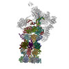 5mp9C 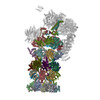 5mpaC 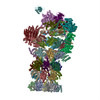 5mpbC 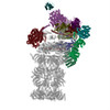 5mpdC 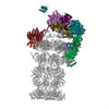 5mpeC M: atomic model generated by this map C: citing same article ( |
|---|---|
| Similar structure data |
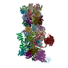
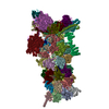
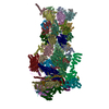
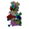
-Links
| EMDB pages | EMDB (EBI/PDBe) / EMDataResource |
|---|---|
| Related items in Molecule of the Month |








-Map
| File | Download / File: emd_3537.map.gz / Format: CCP4 / Size: 274.6 MB / Type: IMAGE STORED AS FLOATING POINT NUMBER (4 BYTES) | ||||||||||||||||||||||||||||||||||||||||||||||||||||||||||||
|---|---|---|---|---|---|---|---|---|---|---|---|---|---|---|---|---|---|---|---|---|---|---|---|---|---|---|---|---|---|---|---|---|---|---|---|---|---|---|---|---|---|---|---|---|---|---|---|---|---|---|---|---|---|---|---|---|---|---|---|---|---|
| Annotation | 26S proteasome in presence of BeFx (s4) | ||||||||||||||||||||||||||||||||||||||||||||||||||||||||||||
| Projections & slices | Image control
Images are generated by Spider. | ||||||||||||||||||||||||||||||||||||||||||||||||||||||||||||
| Voxel size | X=Y=Z: 1.35 Å | ||||||||||||||||||||||||||||||||||||||||||||||||||||||||||||
| Density |
| ||||||||||||||||||||||||||||||||||||||||||||||||||||||||||||
| Symmetry | Space group: 1 | ||||||||||||||||||||||||||||||||||||||||||||||||||||||||||||
| Details | EMDB XML:
CCP4 map header:
| ||||||||||||||||||||||||||||||||||||||||||||||||||||||||||||
 Z (Sec.)
 Y (Row.)
 X (Col.)


-Supplemental data
- Sample components
+Entire : 26S proteasome of Saccharomyces cerevisiae in presence of BeFx (s4)
+Supramolecule #1: 26S proteasome of Saccharomyces cerevisiae in presence of BeFx (s4)
+Macromolecule #1: Proteasome subunit alpha type-1
+Macromolecule #2: Proteasome subunit alpha type-2
+Macromolecule #3: Proteasome subunit alpha type-3
+Macromolecule #4: Proteasome subunit alpha type-4
+Macromolecule #5: Proteasome subunit alpha type-5
+Macromolecule #6: Proteasome subunit alpha type-6
+Macromolecule #7: Probable proteasome subunit alpha type-7
+Macromolecule #8: Proteasome subunit beta type-1
+Macromolecule #9: Proteasome subunit beta type-2
+Macromolecule #10: Proteasome subunit beta type-3
+Macromolecule #11: Proteasome subunit beta type-4
+Macromolecule #12: Proteasome subunit beta type-5
+Macromolecule #13: Proteasome subunit beta type-6
+Macromolecule #14: Proteasome subunit beta type-7
+Macromolecule #15: 26S protease regulatory subunit 7 homolog
+Macromolecule #16: 26S protease regulatory subunit 4 homolog
+Macromolecule #17: 26S protease regulatory subunit 6B homolog
+Macromolecule #18: 26S protease subunit RPT4
+Macromolecule #19: 26S protease regulatory subunit 6A
+Macromolecule #20: 26S protease regulatory subunit 8 homolog
+Macromolecule #21: 26S proteasome regulatory subunit RPN10
+Macromolecule #22: Ubiquitin carboxyl-terminal hydrolase RPN11
+Macromolecule #23: 26S proteasome regulatory subunit RPN12
+Macromolecule #24: 26S proteasome regulatory subunit RPN13
+Macromolecule #25: 26S proteasome complex subunit SEM1
+Macromolecule #26: 26S proteasome regulatory subunit RPN1
+Macromolecule #27: 26S proteasome regulatory subunit RPN2
+Macromolecule #28: 26S proteasome regulatory subunit RPN3
+Macromolecule #29: 26S proteasome regulatory subunit RPN5
+Macromolecule #30: 26S proteasome regulatory subunit RPN6
+Macromolecule #31: 26S proteasome regulatory subunit RPN7
+Macromolecule #32: 26S proteasome regulatory subunit RPN8
+Macromolecule #33: 26S proteasome regulatory subunit RPN9
+Macromolecule #34: Ubiquitin carboxyl-terminal hydrolase 6
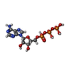
+Macromolecule #35: ADENOSINE-5'-TRIPHOSPHATE
+Macromolecule #36: MAGNESIUM ION
+Macromolecule #37: ADENOSINE-5'-DIPHOSPHATE

-Experimental details
-Structure determination
| Method | cryo EM |
|---|---|
 Processing Processing | single particle reconstruction |
| Aggregation state | particle |
-Sample preparation
| Concentration | 1 mg/mL |
|---|---|
| Buffer | pH: 7.4 |
| Grid | Model: Quantifoil R2/1 / Material: COPPER / Mesh: 400 / Pretreatment - Type: GLOW DISCHARGE / Pretreatment - Time: 15 sec. |
| Vitrification | Cryogen name: ETHANE-PROPANE / Chamber humidity: 95 % / Chamber temperature: 277 K / Instrument: FEI VITROBOT MARK IV |
- Electron microscopy
| Microscope | FEI TITAN KRIOS |
|---|---|
| Image recording | Film or detector model: FEI FALCON II (4k x 4k) / Average electron dose: 45.0 e/Å2 |
| Electron beam | Acceleration voltage: 300 kV / Electron source:  FIELD EMISSION GUN FIELD EMISSION GUN |
| Electron optics | Illumination mode: FLOOD BEAM / Imaging mode: BRIGHT FIELD / Nominal defocus max: 3.5 µm / Nominal defocus min: 1.5 µm |
| Experimental equipment |  Model: Titan Krios / Image courtesy: FEI Company |
-Image processing
| Startup model | Type of model: PDB ENTRY PDB model - PDB ID: |
|---|---|
| Final reconstruction | Applied symmetry - Point group: C1 (asymmetric) / Resolution.type: BY AUTHOR / Resolution: 7.7 Å / Resolution method: FSC 0.143 CUT-OFF / Software - Name: RELION (ver. 1.4) / Number images used: 27600 |
| Initial angle assignment | Type: PROJECTION MATCHING / Software - Name: RELION (ver. 1.4) |
| Final angle assignment | Type: PROJECTION MATCHING / Software - Name: RELION (ver. 1.4) |