 Movie
Movie Controller


+ Open data

- Basic information
| Entry | 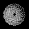 | |||||||||
|---|---|---|---|---|---|---|---|---|---|---|
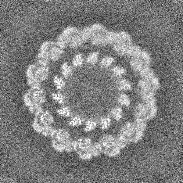
| Title | structure of vp51 in white spot syndrome virus capsid | |||||||||
 Map data Map data | ||||||||||
 Sample Sample |
| |||||||||
 Keywords Keywords | Complex / C14 / capsid / disc / VIRAL PROTEIN | |||||||||
| Function / homology | viral envelope / DNA binding / Capsid protein Function and homology information Function and homology information | |||||||||
| Biological species |  White spot syndrome virus White spot syndrome virus | |||||||||
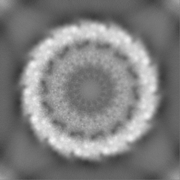
| Method | single particle reconstruction / cryo EM / negative staining / Resolution: 3.73 Å | |||||||||
 Authors Authors | Shan H / Liu MD / Shen QT | |||||||||
| Funding support |  China, 1 items China, 1 items
| |||||||||
 Citation Citation | Journal: To Be Published Title: structure of vp51 in white spot syndrome virus capsid Authors: Hong S / Shen QT | |||||||||
| History |
|
- Structure visualization
| Supplemental images |
|---|
- Downloads & links
-EMDB archive
| Map data | emd_33168.map.gz | 408 MB | EMDB map data format | |
|---|---|---|---|---|
| Header (meta data) | emd-33168-v30.xmlemd-33168.xml | 12.9 KB 12.9 KB | Display Display | EMDB header |
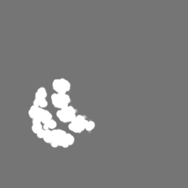
| Images |  emd_33168.png emd_33168.png | 143.9 KB | ||
| Masks | emd_33168_msk_1.map | 824 MB | Mask map | |
| Filedesc metadata | emd-33168.cif.gz | 5 KB | ||
| Others | emd_33168_half_map_1.map.gzemd_33168_half_map_2.map.gz | 764.6 MB 764.6 MB | ||
| Archive directory |  http://ftp.pdbj.org/pub/emdb/structures/EMD-33168ftp://ftp.pdbj.org/pub/emdb/structures/EMD-33168 http://ftp.pdbj.org/pub/emdb/structures/EMD-33168ftp://ftp.pdbj.org/pub/emdb/structures/EMD-33168 | HTTPS FTP |
-Related structure data
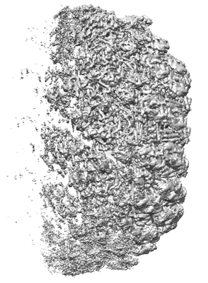
| Related structure data |  7xf2MC M: atomic model generated by this map C: citing same article ( |
|---|---|
| Similar structure data |
-Links
| EMDB pages | EMDB (EBI/PDBe) / EMDataResource |
|---|
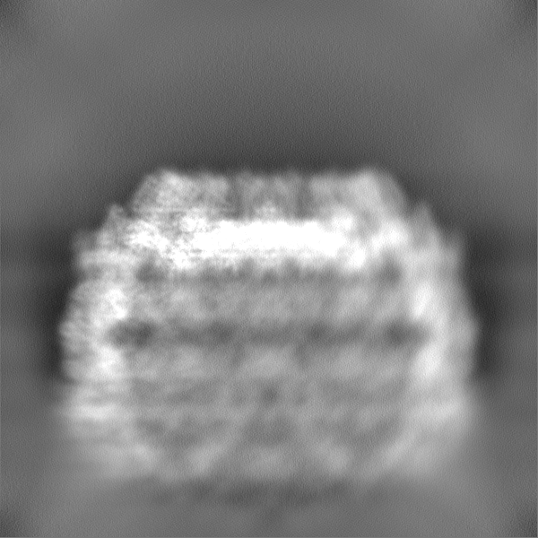
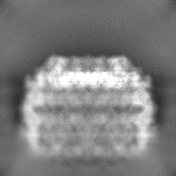
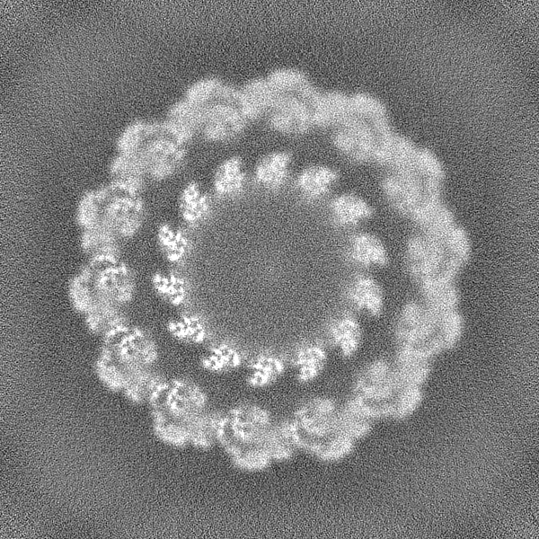
-Map
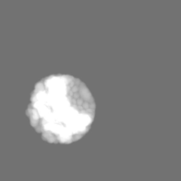
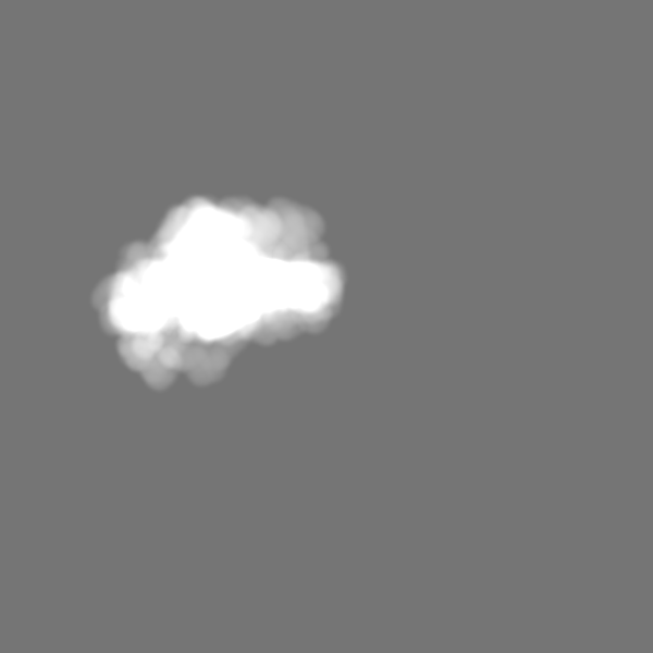
| File | Download / File: emd_33168.map.gz / Format: CCP4 / Size: 824 MB / Type: IMAGE STORED AS FLOATING POINT NUMBER (4 BYTES) | ||||||||||||||||||||||||||||||||||||
|---|---|---|---|---|---|---|---|---|---|---|---|---|---|---|---|---|---|---|---|---|---|---|---|---|---|---|---|---|---|---|---|---|---|---|---|---|---|
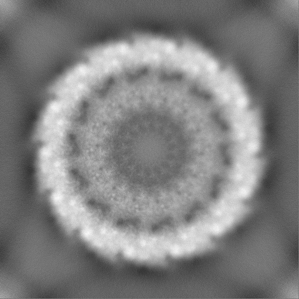
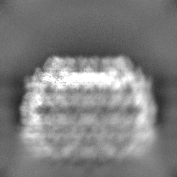
| Projections & slices | Image control
Images are generated by Spider. | ||||||||||||||||||||||||||||||||||||
| Voxel size | X=Y=Z: 1.34 Å | ||||||||||||||||||||||||||||||||||||
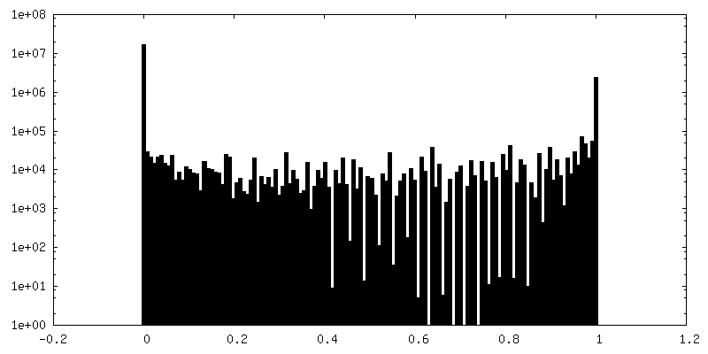
| Density |
| ||||||||||||||||||||||||||||||||||||
| Symmetry | Space group: 1 | ||||||||||||||||||||||||||||||||||||
| Details | EMDB XML:
|
 Z (Sec.)
 Y (Row.)
 X (Col.)


-Supplemental data
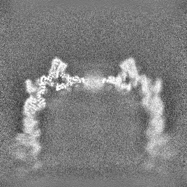
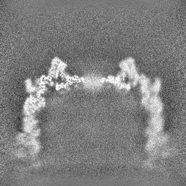
-Mask #1
| File | emd_33168_msk_1.map | ||||||||||||
|---|---|---|---|---|---|---|---|---|---|---|---|---|---|
| Projections & Slices |
| ||||||||||||
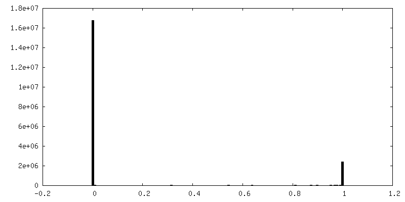
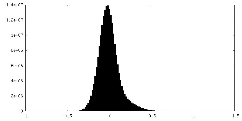
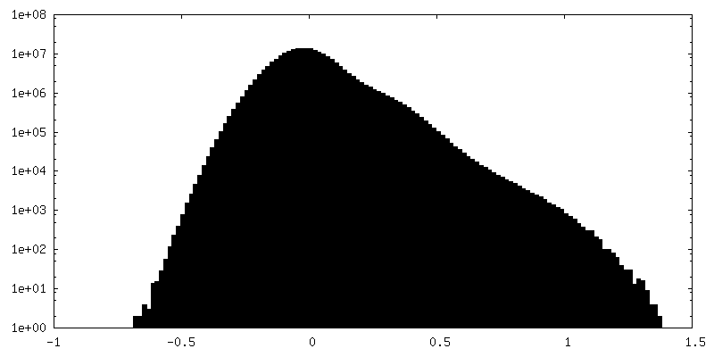
| Density Histograms |


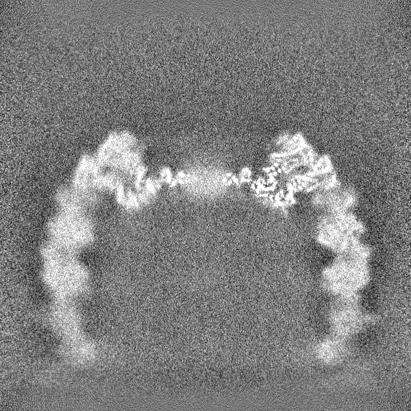
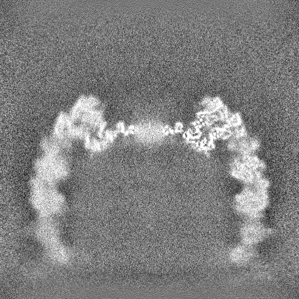
-Half map: #2
| File | emd_33168_half_map_1.map | ||||||||||||
|---|---|---|---|---|---|---|---|---|---|---|---|---|---|
| Projections & Slices |
| ||||||||||||
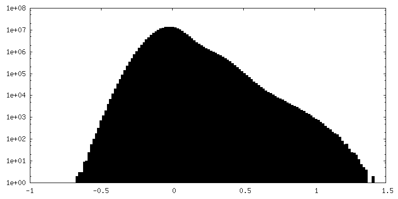
| Density Histograms |

-Half map: #1
| File | emd_33168_half_map_2.map | ||||||||||||
|---|---|---|---|---|---|---|---|---|---|---|---|---|---|
| Projections & Slices |
| ||||||||||||
| Density Histograms |

- Sample components
-Entire : white spot syndrome virus
| Entire | Name: white spot syndrome virus |
|---|---|
| Components |
|
-Supramolecule #1: white spot syndrome virus
| Supramolecule | Name: white spot syndrome virus / type: complex / ID: 1 / Parent: 0 / Macromolecule list: all |
|---|---|
| Source (natural) | Organism: White spot syndrome virus |
-Macromolecule #1: Capsid protein
| Macromolecule | Name: Capsid protein / type: protein_or_peptide / ID: 1 / Number of copies: 2 / Enantiomer: LEVO |
|---|---|
| Source (natural) | Organism: White spot syndrome virus |
| Molecular weight | Theoretical: 51.965145 KDa |
| Sequence | String: MSASLILDEY LKKTASAVLD VADSFEKIKG EIQSPEEAAA LSVALYGAPP KPSASAVASI ITGERTSLND KYLSDNVLLK MSVARVGQE NNRKRADQAA DEIRTIMEDI TGSLSGAYRQ YSPLEEENKV HIGIMNNKTP SIVCGYYTMD TSISSEPLSL T DFQNPTVI ...String: MSASLILDEY LKKTASAVLD VADSFEKIKG EIQSPEEAAA LSVALYGAPP KPSASAVASI ITGERTSLND KYLSDNVLLK MSVARVGQE NNRKRADQAA DEIRTIMEDI TGSLSGAYRQ YSPLEEENKV HIGIMNNKTP SIVCGYYTMD TSISSEPLSL T DFQNPTVI ANVTKRMESI FSKVDSARST RFDAFVNGVA NNMDIKSSID WANMVENVIK LPDSTPNPCS VDTIVSRDAS VV KTAVNDI YASVGKSYCR PATQLTFMSE IEKLRKAAVV CFEALMSDTR ERAFVEFLFY VSFKEDASNT NSKLFVQNKL SSM SGNPRQ PIKLVRRSAE ETLFGLCFMF KVMPPEFMNC IFNFPTIPHS TQYHGLYGTC LTPLLRKYGS SFEKSWAHFE EILS ERANA VKKFGVNDTR IDCLDAVANL TGPVYVLILD LVRTLSAQRS CSTKFLREIK ENYLLWNRFV S UniProtKB: Capsid protein |
-Experimental details
-Structure determination
| Method | negative staining, cryo EM |
|---|---|
 Processing Processing | single particle reconstruction |
| Aggregation state | particle |
-Sample preparation
| Buffer | pH: 8 |
|---|---|
| Staining | Type: NEGATIVE / Material: uranyl acetate |
| Vitrification | Cryogen name: ETHANE |
- Electron microscopy
| Microscope | FEI TITAN KRIOS |
|---|---|
| Image recording | Film or detector model: GATAN K3 BIOQUANTUM (6k x 4k) / Average electron dose: 1.25 e/Å2 |
| Electron beam | Acceleration voltage: 300 kV / Electron source:  FIELD EMISSION GUN FIELD EMISSION GUN |
| Electron optics | C2 aperture diameter: 50.0 µm / Illumination mode: FLOOD BEAM / Imaging mode: BRIGHT FIELD / Cs: 2.7 mm / Nominal defocus max: 1.8 µm / Nominal defocus min: 1.0 µm |
| Experimental equipment |  Model: Titan Krios / Image courtesy: FEI Company |
-Image processing
| Startup model | Type of model: OTHER |
|---|---|
| Final reconstruction | Resolution.type: BY AUTHOR / Resolution: 3.73 Å / Resolution method: FSC 0.143 CUT-OFF / Number images used: 153916 |
| Initial angle assignment | Type: PROJECTION MATCHING |
| Final angle assignment | Type: PROJECTION MATCHING |
