 Movie
Movie Controller

[English] 日本語
 Yorodumi
- PDB-2nsu: Crystal structure of the ectodomain of human transferrin receptor... -
+ Open data

- Basic information
| Entry | Database: PDB / ID: 2nsu | ||||||
|---|---|---|---|---|---|---|---|
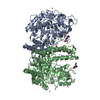
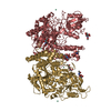
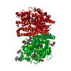
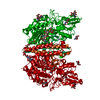
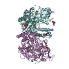
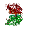
| Title | Crystal structure of the ectodomain of human transferrin receptor fitted into a cryo-EM reconstruction of canine parvovirus and feline transferrin receptor complex | ||||||
 Components Components | Transferrin receptor protein 1 | ||||||
 Keywords Keywords | METAL TRANSPORT / transferrin receptor / virus-receptor complex | ||||||
| Function / homology |  Function and homology information Function and homology informationtransferrin receptor activity / postsynaptic recycling endosome membrane / negative regulation of mitochondrial fusion / positive regulation of isotype switching / Transferrin endocytosis and recycling / response to manganese ion / Differentiation of Keratinocytes in Interfollicular Epidermis in Mammalian Skin / RND1 GTPase cycle / response to iron ion / RND2 GTPase cycle ...transferrin receptor activity / postsynaptic recycling endosome membrane / negative regulation of mitochondrial fusion / positive regulation of isotype switching / Transferrin endocytosis and recycling / response to manganese ion / Differentiation of Keratinocytes in Interfollicular Epidermis in Mammalian Skin / RND1 GTPase cycle / response to iron ion / RND2 GTPase cycle / RHOB GTPase cycle / response to copper ion / RHOJ GTPase cycle / RHOC GTPase cycle / Golgi Associated Vesicle Biogenesis / RHOQ GTPase cycle / CDC42 GTPase cycle / RHOG GTPase cycle / RHOH GTPase cycle / RAC3 GTPase cycle / RHOA GTPase cycle / RAC2 GTPase cycle / response to retinoic acid / regulation of postsynaptic membrane neurotransmitter receptor levels / transport across blood-brain barrier / positive regulation of B cell proliferation / RAC1 GTPase cycle / clathrin-coated pit / response to nutrient / positive regulation of T cell proliferation / osteoclast differentiation / Hsp70 protein binding / receptor-mediated endocytosis / acute-phase response / cellular response to leukemia inhibitory factor / iron ion transport / clathrin-coated endocytic vesicle membrane / HFE-transferrin receptor complex / transferrin transport / positive regulation of protein-containing complex assembly / multicellular organismal-level iron ion homeostasis / receptor internalization / positive regulation of protein localization to nucleus / recycling endosome / cellular response to xenobiotic stimulus / recycling endosome membrane / melanosome / Cargo recognition for clathrin-mediated endocytosis / double-stranded RNA binding / extracellular vesicle / Clathrin-mediated endocytosis / virus receptor activity / cytoplasmic vesicle / blood microparticle / basolateral plasma membrane / intracellular iron ion homeostasis / response to hypoxia / early endosome / positive regulation of canonical NF-kappaB signal transduction / cell surface receptor signaling pathway / endosome / endosome membrane / intracellular signal transduction / external side of plasma membrane / positive regulation of gene expression / protein kinase binding / negative regulation of apoptotic process / protein-containing complex binding / perinuclear region of cytoplasm / glutamatergic synapse / cell surface / protein homodimerization activity / : / RNA binding / extracellular exosome / extracellular region / membrane / identical protein binding / plasma membrane Similarity search - Function | ||||||
| Biological species |  Homo sapiens (human) Homo sapiens (human) | ||||||
| Method | ELECTRON MICROSCOPY / single particle reconstruction / cryo EM / Resolution: 27 Å | ||||||
 Authors Authors | Hafenstein, S. / Kostyuchenko, V.A. / Rossmann, M.G. | ||||||
 Citation Citation | Journal: Proc Natl Acad Sci U S A / Year: 2007 Title: Asymmetric binding of transferrin receptor to parvovirus capsids. Authors: Susan Hafenstein / Laura M Palermo / Victor A Kostyuchenko / Chuan Xiao / Marc C Morais / Christian D S Nelson / Valorie D Bowman / Anthony J Battisti / Paul R Chipman / Colin R Parrish / Michael G Rossmann /  Abstract: Although many viruses are icosahedral when they initially bind to one or more receptor molecules on the cell surface, such an interaction is asymmetric, probably causing a breakdown in the symmetry ...Although many viruses are icosahedral when they initially bind to one or more receptor molecules on the cell surface, such an interaction is asymmetric, probably causing a breakdown in the symmetry and conformation of the original infecting virion in preparation for membrane penetration and release of the viral genome. Cryoelectron microscopy and biochemical analyses show that transferrin receptor, the cellular receptor for canine parvovirus, can bind to only one or a few of the 60 icosahedrally equivalent sites on the virion, indicating that either canine parvovirus has inherent asymmetry or binding of receptor induces asymmetry. The asymmetry of receptor binding to canine parvovirus is reminiscent of the special portal in tailed bacteriophages and some large, icosahedral viruses. Asymmetric interactions of icosahedral viruses with their hosts might be a more common phenomenon than previously thought and may have been obscured by averaging in previous crystallographic and electron microscopic structure determinations. | ||||||
| History |
| ||||||
| Remark 999 | SEQUENCE AUTHORS STATE THAT PDB ENTRY 1CX8 WAS USED FOR FITTING IN THIS ENTRY. THE SEQUENCE ... SEQUENCE AUTHORS STATE THAT PDB ENTRY 1CX8 WAS USED FOR FITTING IN THIS ENTRY. THE SEQUENCE DIFFERENCES EXIST IN THE STRUCTURE 1CX8. THE UNIPROT ENTRY P02786 IS A RESULT OF DNA SEQUENCING, WHILE 1CX8 SEQUENCE IS APPARENTLY BASED ON MRNA SEQUENCE. |
- Structure visualization
| Movie |
Movie viewer |
|---|---|
| Structure viewer | Molecule: MolmilJmol/JSmol |
- Downloads & links
-Download
| PDBx/mmCIF format | 2nsu.cif.gz | 253.2 KB | Display | PDBx/mmCIF format |
|---|---|---|---|---|
| PDB format | pdb2nsu.ent.gz | 203 KB | Display | PDB format |
| PDBx/mmJSON format | 2nsu.json.gz | Tree view | PDBx/mmJSON format | |
| Others |  Other downloads Other downloads |
-Validation report
| Arichive directory | https://data.pdbj.org/pub/pdb/validation_reports/ns/2nsuftp://data.pdbj.org/pub/pdb/validation_reports/ns/2nsu | HTTPS FTP |
|---|
-Related structure data
| Related structure data | 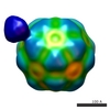 1288MC 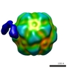 1287C M: map data used to model this data C: citing same article ( |
|---|---|
| Similar structure data |


-Links
 PDBj
PDBj


- Assembly
| Deposited unit | 
|
|---|---|
| 1 |
|
-Components
| #1: Protein | Mass: 71622.961 Da / Num. of mol.: 2 / Fragment: THE ECTODOMAIN OF HUMAN TRANSFERRIN RECEPTOR Source method: isolated from a genetically manipulated source Source: (gene. exp.) Homo sapiens (human) / Gene: TFRC / Plasmid: PCMVTFR / Organ (production host): OVARY CELLS / Production host:   Cricetulus griseus (Chinese hamster) / References: UniProt: P02786 Cricetulus griseus (Chinese hamster) / References: UniProt: P02786Has protein modification | Y | |
|---|
-Experimental details
-Experiment
| Experiment | Method: ELECTRON MICROSCOPY |
|---|---|
| EM experiment | Aggregation state: PARTICLE / 3D reconstruction method: single particle reconstruction |
- Sample preparation
| Component | Name: MIXTURE OF EMPTY CANINE PARVOVIRUS CAPSIDS AND ECTODOMAINS OF FELINE TRANSFERRIN RECEPTORS Type: VIRUS / Details: Based on PDB entry 1CX8 |
|---|---|
| Buffer solution | Name: 0.02M TRIS-HCL / pH: 7.5 / Details: 0.02M TRIS-HCL |
| Specimen | Embedding applied: NO / Shadowing applied: NO / Staining applied: NO / Vitrification applied: YES |
| Specimen support | Details: QUANTIFOIL 200 |
| Vitrification | Cryogen name: ETHANE / Details: PLUNGED IN LIQUID ETHANE |
- Electron microscopy imaging
| Microscopy | Model: FEI/PHILIPS CM200FEG / Date: Jan 22, 2003 |
|---|---|
| Electron gun | Electron source:  FIELD EMISSION GUN / Accelerating voltage: 200 kV / Illumination mode: FLOOD BEAM FIELD EMISSION GUN / Accelerating voltage: 200 kV / Illumination mode: FLOOD BEAM |
| Electron lens | Mode: BRIGHT FIELD / Nominal magnification: 50000 X / Calibrated magnification: 54000 X / Nominal defocus max: 3900 nm / Nominal defocus min: 1700 nm / Cs: 2 mm |
| Image recording | Electron dose: 25.96 e/Å2 / Film or detector model: KODAK SO-163 FILM |
| Image scans | Num. digital images: 115 |
| Radiation | Protocol: SINGLE WAVELENGTH / Monochromatic (M) / Laue (L): M |
| Radiation wavelength | Relative weight: 1 |
- Processing
| EM software |
| ||||||||||||
|---|---|---|---|---|---|---|---|---|---|---|---|---|---|
| CTF correction | Details: CTF correction of each particle | ||||||||||||
| Symmetry | Point symmetry: C2 (2 fold cyclic) | ||||||||||||
| 3D reconstruction | Method: projection matching / Resolution: 27 Å / Num. of particles: 8566 / Nominal pixel size: 2.6 Å / Actual pixel size: 2.6 Å / Details: a modified version of XMIPP software was used / Symmetry type: POINT | ||||||||||||
| Atomic model building | Protocol: RIGID BODY FIT / Space: REAL / Target criteria: BEST VISUAL FIT USING THE PROGRAM O Details: METHOD--MANUAL FITTING USING THE PROGRAM O REFINEMENT PROTOCOL--RIGID BODY | ||||||||||||
| Atomic model building | PDB-ID: 1CX8 Accession code: 1CX8 / Source name: PDB / Type: experimental model | ||||||||||||
| Refinement | Highest resolution: 27 Å | ||||||||||||
| Refinement step | Cycle: LAST / Highest resolution: 27 Å
|
