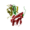
[pyruvate dehydrogenase (acetyl-transferring)] kinase / regulation of fatty acid oxidation / pyruvate dehydrogenase (acetyl-transferring) kinase activity / regulation of pyruvate decarboxylation to acetyl-CoA / Regulation of pyruvate dehydrogenase (PDH) complex / regulation of fatty acid biosynthetic process / regulation of ketone metabolic process / regulation of pH / cellular response to fatty acid / regulation of bone resorption ...[pyruvate dehydrogenase (acetyl-transferring)] kinase / regulation of fatty acid oxidation / pyruvate dehydrogenase (acetyl-transferring) kinase activity / regulation of pyruvate decarboxylation to acetyl-CoA / Regulation of pyruvate dehydrogenase (PDH) complex / regulation of fatty acid biosynthetic process / regulation of ketone metabolic process / regulation of pH / cellular response to fatty acid / regulation of bone resorption / Signaling by Retinoic Acid / response to starvation / regulation of glucose metabolic process / cellular response to starvation / insulin receptor signaling pathway / glucose homeostasis / MLL4 and MLL3 complexes regulate expression of PPARG target genes in adipogenesis and hepatic steatosis / protein kinase activity / mitochondrial matrix / mitochondrion / ATP binding Similarity search - Function
In the structure databanks used in Yorodumi, some data are registered as the other names, "COVID-19 virus" and "2019-nCoV". Here are the details of the virus and the list of structure data.
Jan 31, 2019. EMDB accession codes are about to change! (news from PDBe EMDB page)
EMDB accession codes are about to change! (news from PDBe EMDB page)
The allocation of 4 digits for EMDB accession codes will soon come to an end. Whilst these codes will remain in use, new EMDB accession codes will include an additional digit and will expand incrementally as the available range of codes is exhausted. The current 4-digit format prefixed with “EMD-” (i.e. EMD-XXXX) will advance to a 5-digit format (i.e. EMD-XXXXX), and so on. It is currently estimated that the 4-digit codes will be depleted around Spring 2019, at which point the 5-digit format will come into force.
The EM Navigator/Yorodumi systems omit the EMD- prefix.
Related info.:Q: What is EMD? / ID/Accession-code notation in Yorodumi/EM Navigator
Yorodumi is a browser for structure data from EMDB, PDB, SASBDB, etc.
This page is also the successor to EM Navigator detail page, and also detail information page/front-end page for Omokage search.
The word "yorodu" (or yorozu) is an old Japanese word meaning "ten thousand". "mi" (miru) is to see.
Related info.:EMDB / PDB / SASBDB / Comparison of 3 databanks / Yorodumi Search / Aug 31, 2016. New EM Navigator & Yorodumi / Yorodumi Papers / Jmol/JSmol / Function and homology information / Changes in new EM Navigator and Yorodumi
 Movie
Movie Controller

 Yorodumi
 Open data
 Basic information
 Components
 Keywords
 Function and homology information
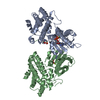
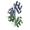
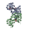
 Homo sapiens (human)
 X-RAY DIFFRACTION /
  Authors
 Citation
 Structure visualization
 Downloads & links
 Other downloads


 PDBj
PDBj


 Assembly
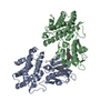

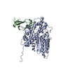
 Spodoptera frugiperda (fall armyworm)
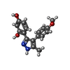
 Mass: 296.321 Da / Num. of mol.: 2 / Source method: obtained synthetically / Formula: C17H16N2O3
 Mass: 18.015 Da / Num. of mol.: 79 / Source method: isolated from a natural source / Formula: H2O
 Sample preparation
 / Beamline: BL32B2 / Wavelength: 1 Å
 Processing