[English] 日本語
 Yorodumi
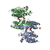
- PDB-2gms: E coli GDP-4-keto-6-deoxy-D-mannose-3-dehydratase with bound hydr... -
+ Open data
- Basic information
| Entry | Database: PDB / ID: 2gms | ||||||
|---|---|---|---|---|---|---|---|
| Title | E coli GDP-4-keto-6-deoxy-D-mannose-3-dehydratase with bound hydrated PLP | ||||||
 Components Components | Putative pyridoxamine 5-phosphate-dependent dehydrase, Wbdk | ||||||
 Keywords Keywords | TRANSFERASE / colitose / 0-antigen / aspartate aminotransferase / plp / deoxysugar | ||||||
| Function / homology |  Function and homology information Function and homology informationTransferases; Transferring nitrogenous groups; Transaminases / polysaccharide biosynthetic process / transaminase activity / pyridoxal phosphate binding / metal ion binding Similarity search - Function | ||||||
| Biological species |  | ||||||
| Method |  X-RAY DIFFRACTION / X-RAY DIFFRACTION /  MOLECULAR REPLACEMENT / Resolution: 1.8 Å MOLECULAR REPLACEMENT / Resolution: 1.8 Å | ||||||
 Authors Authors | Cook, P.D. / Thoden, J.B. / Holden, H.M. | ||||||
 Citation Citation |  Journal: Protein Sci. / Year: 2006 Journal: Protein Sci. / Year: 2006Title: The structure of GDP-4-keto-6-deoxy-D-mannose-3-dehydratase: a unique coenzyme B6-dependent enzyme. Authors: Cook, P.D. / Thoden, J.B. / Holden, H.M. | ||||||
| History |
|
- Structure visualization
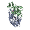
| Structure viewer | Molecule:  Molmil Molmil Jmol/JSmol Jmol/JSmol |
|---|
- Downloads & links
- Download
| PDBx/mmCIF format |  2gms.cif.gz 2gms.cif.gz | 166 KB | Display |  PDBx/mmCIF format PDBx/mmCIF format |
|---|---|---|---|---|
| PDB format |  pdb2gms.ent.gz pdb2gms.ent.gz | 130.1 KB | Display |  PDB format PDB format |
| PDBx/mmJSON format |  2gms.json.gz 2gms.json.gz | Tree view |  PDBx/mmJSON format PDBx/mmJSON format | |
| Others |  Other downloads Other downloads |
-Validation report
| Summary document |  2gms_validation.pdf.gz 2gms_validation.pdf.gz | 1.1 MB | Display |  wwPDB validaton report wwPDB validaton report |
|---|---|---|---|---|
| Full document |  2gms_full_validation.pdf.gz 2gms_full_validation.pdf.gz | 1.1 MB | Display | |
| Data in XML |  2gms_validation.xml.gz 2gms_validation.xml.gz | 30.4 KB | Display | |
| Data in CIF |  2gms_validation.cif.gz 2gms_validation.cif.gz | 41.7 KB | Display | |
| Arichive directory |  https://data.pdbj.org/pub/pdb/validation_reports/gm/2gms https://data.pdbj.org/pub/pdb/validation_reports/gm/2gms ftp://data.pdbj.org/pub/pdb/validation_reports/gm/2gms ftp://data.pdbj.org/pub/pdb/validation_reports/gm/2gms | HTTPS FTP |
-Related structure data
| Related structure data | 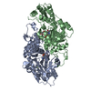 2gmuC  1mdoS C: citing same article ( S: Starting model for refinement |
|---|---|
| Similar structure data |
- Links
Links
- Assembly
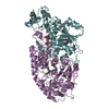
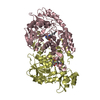
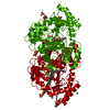
| Deposited unit | 
| ||||||||
|---|---|---|---|---|---|---|---|---|---|
| 1 |
| ||||||||
| Unit cell |
|
- Components
| #1: Protein | Mass: 44352.422 Da / Num. of mol.: 2 Source method: isolated from a genetically manipulated source Source: (gene. exp.)   #2: Chemical | ChemComp-MG / | #3: Chemical | #4: Water | ChemComp-HOH / | |
|---|
-Experimental details
-Experiment
| Experiment | Method:  X-RAY DIFFRACTION / Number of used crystals: 1 X-RAY DIFFRACTION / Number of used crystals: 1 |
|---|
- Sample preparation
| Crystal | Density Matthews: 2.34 Å3/Da / Density % sol: 47.41 % |
|---|---|
| Crystal grow | Temperature: 298 K / pH: 6 Details: 14% PEG 3400, 0.5 M MES buffer, 0.2 magnesium chloride, 0.1 M sodium chloride, 1 mM pyridoxal-5-phosphate, 2 mM 2-ketoglutarate, 1 mM GDP, Batch, pH 6.0, temperature 298K |
-Data collection
| Diffraction | Mean temperature: 277 K |
|---|---|
| Diffraction source | Source:  ROTATING ANODE / Type: RIGAKU RU200 / Wavelength: 1.5418 Å ROTATING ANODE / Type: RIGAKU RU200 / Wavelength: 1.5418 Å |
| Detector | Type: SIEMENS HI-STAR / Detector: AREA DETECTOR / Date: Oct 12, 2005 / Details: supper mirrors |
| Radiation | Monochromator: ni filter / Protocol: SINGLE WAVELENGTH / Monochromatic (M) / Laue (L): M / Scattering type: x-ray |
| Radiation wavelength | Wavelength: 1.5418 Å / Relative weight: 1 |
| Reflection | Resolution: 1.8→30 Å / Num. all: 76612 / Num. obs: 76612 / % possible obs: 92 % / Observed criterion σ(F): 0 / Observed criterion σ(I): 0 / Redundancy: 3.7 % / Rsym value: 0.0586 / Net I/σ(I): 9.9 |
| Reflection shell | Resolution: 1.8→1.88 Å / Redundancy: 1.5 % / Mean I/σ(I) obs: 1.4 / Num. unique all: 8943 / Rsym value: 0.323 / % possible all: 85 |
- Processing
| Software |
| |||||||||||||||||||||||||
|---|---|---|---|---|---|---|---|---|---|---|---|---|---|---|---|---|---|---|---|---|---|---|---|---|---|---|
| Refinement | Method to determine structure:  MOLECULAR REPLACEMENT MOLECULAR REPLACEMENTStarting model: PDB entry 1MDO Resolution: 1.8→30 Å / Cross valid method: THROUGHOUT / σ(F): 0 / σ(I): 0 / Stereochemistry target values: Engh & Huber
| |||||||||||||||||||||||||
| Refinement step | Cycle: LAST / Resolution: 1.8→30 Å
| |||||||||||||||||||||||||
| Refine LS restraints |
|
 Movie
Movie Controller





 PDBj
PDBj

