 Movie
Movie Controller

[English] 日本語
 Yorodumi
- PDB-2apc: Crystal Structure of N-Acetylglucosaminyltransferase I in Complex... -
+ Open data

- Basic information
| Entry | Database: PDB / ID: 2apc | ||||||
|---|---|---|---|---|---|---|---|
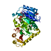
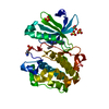
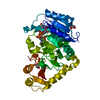
| Title | Crystal Structure of N-Acetylglucosaminyltransferase I in Complex with UDP-GlcNAc phosphonate | ||||||
 Components Components | Alpha-1,3-mannosyl-glycoprotein 2-beta-N-acetylglucosaminyltransferase | ||||||
 Keywords Keywords | TRANSFERASE / N-Acetylglucosaminyltransferase / glycosyltransferase / UDP-GlcNAc / UDP-GlcNAc phosphonate | ||||||
| Function / homology |  Function and homology information Function and homology informationalpha-1,3-mannosyl-glycoprotein 2-beta-N-acetylglucosaminyltransferase / alpha-1,3-mannosylglycoprotein 2-beta-N-acetylglucosaminyltransferase activity / protein N-acetylglucosaminyltransferase activity / mannose metabolic process / : / protein N-linked glycosylation / Golgi medial cisterna / manganese ion binding / in utero embryonic development / Golgi membrane / perinuclear region of cytoplasm Similarity search - Function | ||||||
| Biological species |  | ||||||
| Method |  X-RAY DIFFRACTION / SYNCHROTRON / MOLECULAR REPLACEMENT / Resolution: 1.5 Å X-RAY DIFFRACTION / SYNCHROTRON / MOLECULAR REPLACEMENT / Resolution: 1.5 Å | ||||||
 Authors Authors | Rini, J.M. / Gordon, R.D. | ||||||
 Citation Citation | Journal: J.Mol.Biol. / Year: 2006 Title: X-ray Crystal Structures of Rabbit N-acetylglucosaminyltransferase I (GnT I) in Complex with Donor Substrate Analogues. Authors: Gordon, R.D. / Sivarajah, P. / Satkunarajah, M. / Ma, D. / Tarling, C.A. / Vizitiu, D. / Withers, S.G. / Rini, J.M. | ||||||
| History |
|
- Structure visualization
| Structure viewer | Molecule: MolmilJmol/JSmol |
|---|
- Downloads & links
-Download
| PDBx/mmCIF format | 2apc.cif.gz | 95.3 KB | Display | PDBx/mmCIF format |
|---|---|---|---|---|
| PDB format | pdb2apc.ent.gz | 69.6 KB | Display | PDB format |
| PDBx/mmJSON format | 2apc.json.gz | Tree view | PDBx/mmJSON format | |
| Others |  Other downloads Other downloads |
-Validation report
| Arichive directory | https://data.pdbj.org/pub/pdb/validation_reports/ap/2apcftp://data.pdbj.org/pub/pdb/validation_reports/ap/2apc | HTTPS FTP |
|---|
-Related structure data
| Related structure data | 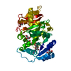 2am3C  2am4C 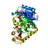 2am5C 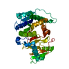 1fo9S  1foaS S: Starting model for refinement C: citing same article ( |
|---|---|
| Similar structure data |


-Links
 PDBj
PDBj


- Assembly
| Deposited unit | 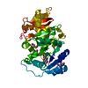
| ||||||||
|---|---|---|---|---|---|---|---|---|---|
| 1 |
| ||||||||
| Unit cell |
|
-Components
| #1: Protein | Mass: 39725.172 Da / Num. of mol.: 1 / Fragment: residues 106-447 Source method: isolated from a genetically manipulated source Source: (gene. exp.)  Cricetulus griseus (Chinese hamster) Cricetulus griseus (Chinese hamster)References: UniProt: P27115, alpha-1,3-mannosyl-glycoprotein 2-beta-N-acetylglucosaminyltransferase | ||||
|---|---|---|---|---|---|
| #2: Chemical | ChemComp-MN /   Mass: 54.938 Da / Num. of mol.: 1 / Source method: obtained synthetically / Formula: Mn Mass: 54.938 Da / Num. of mol.: 1 / Source method: obtained synthetically / Formula: Mn | ||||
| #3: Chemical | ChemComp-UDM /   Mass: 605.381 Da / Num. of mol.: 1 / Source method: obtained synthetically / Formula: C18H29N3O16P2 Mass: 605.381 Da / Num. of mol.: 1 / Source method: obtained synthetically / Formula: C18H29N3O16P2 | ||||
| #4: Chemical | ChemComp-GOL /   Mass: 92.094 Da / Num. of mol.: 6 / Source method: obtained synthetically / Formula: C3H8O3 Mass: 92.094 Da / Num. of mol.: 6 / Source method: obtained synthetically / Formula: C3H8O3#5: Water | ChemComp-HOH / |  Mass: 18.015 Da / Num. of mol.: 278 / Source method: isolated from a natural source / Formula: H2O Mass: 18.015 Da / Num. of mol.: 278 / Source method: isolated from a natural source / Formula: H2OHas protein modification | Y | |
-Experimental details
-Experiment
| Experiment | Method: X-RAY DIFFRACTION / Number of used crystals: 1 |
|---|
- Sample preparation
| Crystal | Density Matthews: 2.1 Å3/Da / Density % sol: 41.04 % |
|---|---|
| Crystal grow | Temperature: 293 K / Method: vapor diffusion, hanging drop / pH: 7.9 Details: 1 M Tris pH 7.9, 40% PEG 6000, MnCl2, VAPOR DIFFUSION, HANGING DROP, temperature 293K |
-Data collection
| Diffraction | Mean temperature: 100 K |
|---|---|
| Diffraction source | Source: SYNCHROTRON / Site: CHESS  / Beamline: A1 / Wavelength: 0.9363 Å / Beamline: A1 / Wavelength: 0.9363 Å |
| Detector | Type: ADSC QUANTUM 210 / Detector: CCD / Date: Feb 3, 2003 |
| Radiation | Monochromator: SYCHROTRON / Protocol: SINGLE WAVELENGTH / Monochromatic (M) / Laue (L): M / Scattering type: x-ray |
| Radiation wavelength | Wavelength: 0.9363 Å / Relative weight: 1 |
| Reflection | Resolution: 1.5→43.51 Å / Num. all: 55630 / Num. obs: 54569 / % possible obs: 96.9 % / Observed criterion σ(F): 0 / Observed criterion σ(I): 0 / Biso Wilson estimate: 10.7 Å2 / Limit h max: 26 / Limit h min: 0 / Limit k max: 54 / Limit k min: 0 / Limit l max: 68 / Limit l min: 0 / Observed criterion F max: 194316.95 / Observed criterion F min: 0.32 |
| Reflection shell | Resolution: 1.5→1.59 Å / % possible all: 86.8 |
- Processing
| Software |
| ||||||||||||||||||||||||||||||||||||||||||||||||||||||||||||||||||||||
|---|---|---|---|---|---|---|---|---|---|---|---|---|---|---|---|---|---|---|---|---|---|---|---|---|---|---|---|---|---|---|---|---|---|---|---|---|---|---|---|---|---|---|---|---|---|---|---|---|---|---|---|---|---|---|---|---|---|---|---|---|---|---|---|---|---|---|---|---|---|---|---|
| Refinement | Method to determine structure: MOLECULAR REPLACEMENT Starting model: 1FOA, 1FO9 Resolution: 1.5→43.51 Å / Rfactor Rfree error: 0.003 / Occupancy max: 1 / Occupancy min: 0.5 / Cross valid method: THROUGHOUT / σ(F): 0 / Stereochemistry target values: Engh & Huber
| ||||||||||||||||||||||||||||||||||||||||||||||||||||||||||||||||||||||
| Solvent computation | Solvent model: CNS bulk solvent model used / Bsol: 35.8371 Å2 / ksol: 0.379612 e/Å3 | ||||||||||||||||||||||||||||||||||||||||||||||||||||||||||||||||||||||
| Displacement parameters | Biso max: 47.62 Å2 / Biso mean: 16.76 Å2 / Biso min: 5.14 Å2 | ||||||||||||||||||||||||||||||||||||||||||||||||||||||||||||||||||||||
| Refine Biso |
| ||||||||||||||||||||||||||||||||||||||||||||||||||||||||||||||||||||||
| Refine analyze |
| ||||||||||||||||||||||||||||||||||||||||||||||||||||||||||||||||||||||
| Refinement step | Cycle: LAST / Resolution: 1.5→43.51 Å
| ||||||||||||||||||||||||||||||||||||||||||||||||||||||||||||||||||||||
| Refine LS restraints |
| ||||||||||||||||||||||||||||||||||||||||||||||||||||||||||||||||||||||
| LS refinement shell | Refine-ID: X-RAY DIFFRACTION
| ||||||||||||||||||||||||||||||||||||||||||||||||||||||||||||||||||||||
| Xplor file |
|
