Movie
Movie Controller

[English] 日本語
 Yorodumi
- EMDB-29807: 70S global refined map of WT E.coli ribosome complexed with A-sit... -
+ Open data

- Basic information
| Entry |  | |||||||||
|---|---|---|---|---|---|---|---|---|---|---|
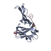
| Title | 70S global refined map of WT E.coli ribosome complexed with A-site 3-aminopyridine-4-carboxylic acid charged tRNAPhe | |||||||||
 Map data Map data | 70S global refined map | |||||||||
 Sample Sample |
| |||||||||
| Biological species |  | |||||||||
| Method | single particle reconstruction / cryo EM / Resolution: 2.09 Å | |||||||||
 Authors Authors | Majumdar C / Cate JHD | |||||||||
| Funding support |  United States, 1 items United States, 1 items
| |||||||||
 Citation Citation | Journal: To Be Published Title: Structure of WT E.coli ribosome 50S subunit with complexed with mRNA, P-site fMet-NH-tRNAfMet and A-site 3-aminopyridine-4-carboxylic acid charged NH-tRNAPhe Authors: Majumdar C / Cate JHD | |||||||||
| History |
|
- Structure visualization
| Supplemental images |
|---|

- Downloads & links
-EMDB archive
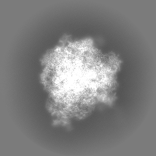
| Map data | emd_29807.map.gz | 55.6 MB |  EMDB map data format EMDB map data format | |
|---|---|---|---|---|
| Header (meta data) | emd-29807-v30.xmlemd-29807.xml | 16.9 KB 16.9 KB | Display Display | EMDB header |
| FSC (resolution estimation) | emd_29807_fsc.xml | 17.4 KB | Display | FSC data file |
| Images |  emd_29807.png emd_29807.png | 82.8 KB | ||
| Others | emd_29807_half_map_1.map.gzemd_29807_half_map_2.map.gz | 372.8 MB 372.8 MB | ||
| Archive directory |  http://ftp.pdbj.org/pub/emdb/structures/EMD-29807ftp://ftp.pdbj.org/pub/emdb/structures/EMD-29807 http://ftp.pdbj.org/pub/emdb/structures/EMD-29807ftp://ftp.pdbj.org/pub/emdb/structures/EMD-29807 | HTTPS FTP |
-Validation report
| Summary document | emd_29807_validation.pdf.gz | 867.2 KB | Display | EMDB validaton report |
|---|---|---|---|---|
| Full document | emd_29807_full_validation.pdf.gz | 866.8 KB | Display | |
| Data in XML | emd_29807_validation.xml.gz | 25.4 KB | Display | |
| Data in CIF | emd_29807_validation.cif.gz | 34 KB | Display | |
| Arichive directory | https://ftp.pdbj.org/pub/emdb/validation_reports/EMD-29807ftp://ftp.pdbj.org/pub/emdb/validation_reports/EMD-29807 | HTTPS FTP |
-Related structure data


-Links
| EMDB pages | EMDB (EBI/PDBe) / EMDataResource |
|---|
-Map
| File | Download / File: emd_29807.map.gz / Format: CCP4 / Size: 465.5 MB / Type: IMAGE STORED AS FLOATING POINT NUMBER (4 BYTES) | ||||||||||||||||||||
|---|---|---|---|---|---|---|---|---|---|---|---|---|---|---|---|---|---|---|---|---|---|
| Annotation | 70S global refined map | ||||||||||||||||||||
| Voxel size | X=Y=Z: 0.824 Å | ||||||||||||||||||||
| Density |
| ||||||||||||||||||||
| Symmetry | Space group: 1 | ||||||||||||||||||||
| Details | EMDB XML:
|
-Supplemental data
-Half map: 70S global refinement half map
| File | emd_29807_half_map_1.map | ||||||||||||
|---|---|---|---|---|---|---|---|---|---|---|---|---|---|
| Annotation | 70S global refinement half map | ||||||||||||
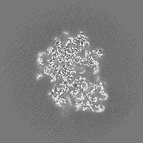
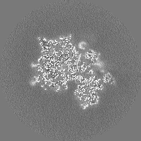
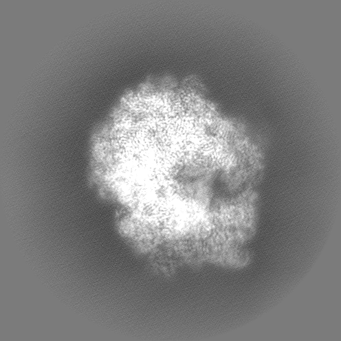
| Projections & Slices |
| ||||||||||||
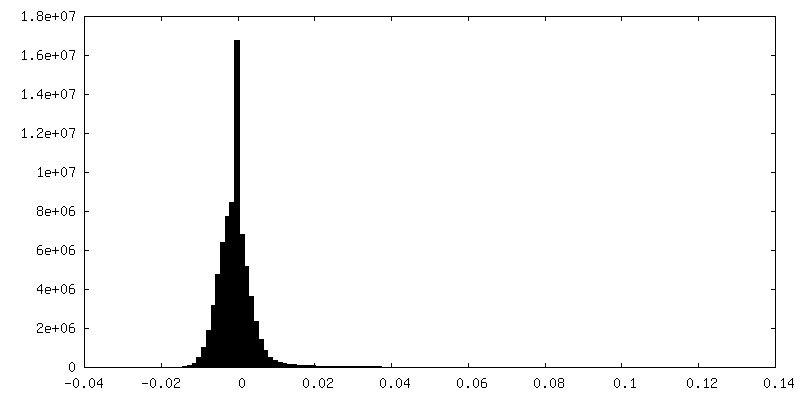
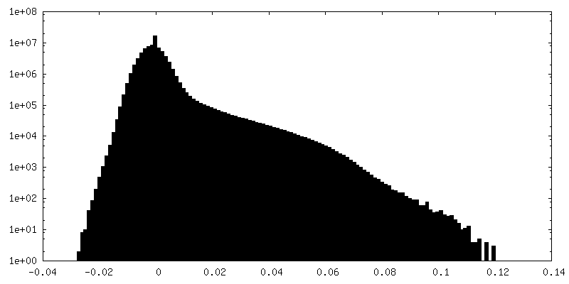
| Density Histograms |
 Z
Z Y
Y X
X

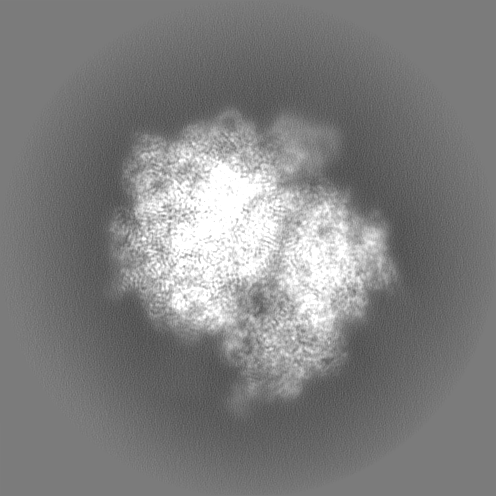

-Half map: 70S global refinement half map
| File | emd_29807_half_map_2.map | ||||||||||||
|---|---|---|---|---|---|---|---|---|---|---|---|---|---|
| Annotation | 70S global refinement half map | ||||||||||||
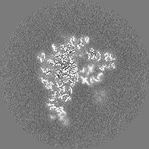
| Projections & Slices |
| ||||||||||||
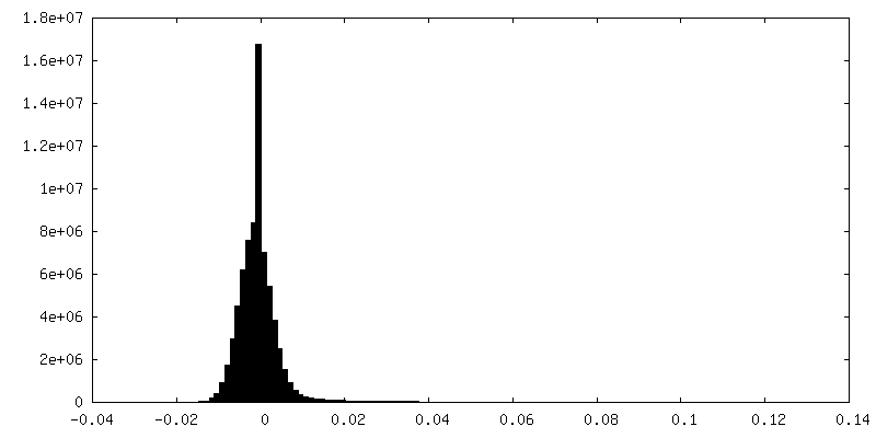
| Density Histograms |


- Sample components
-Entire : 70S E.coli ribosome
| Entire | Name: 70S E.coli ribosome |
|---|---|
| Components |
|
-Supramolecule #1: 70S E.coli ribosome
| Supramolecule | Name: 70S E.coli ribosome / type: complex / ID: 1 / Chimera: Yes / Parent: 0 / Macromolecule list: #1-#34 |
|---|---|
| Source (natural) | Organism: |
-Experimental details
-Structure determination
| Method | cryo EM |
|---|---|
 Processing Processing | single particle reconstruction |
| Aggregation state | particle |
-Sample preparation
| Buffer | pH: 7.5 |
|---|---|
| Grid | Model: Quantifoil R1.2/1.3 / Material: GOLD / Mesh: 300 / Support film - Material: CARBON / Support film - topology: CONTINUOUS / Support film - Film thickness: 0.2 nm / Pretreatment - Type: GLOW DISCHARGE |
| Vitrification | Cryogen name: ETHANE / Chamber humidity: 100 % / Chamber temperature: 277 K / Instrument: FEI VITROBOT MARK IV |
- Electron microscopy
| Microscope | FEI TITAN KRIOS |
|---|---|
| Image recording | Film or detector model: GATAN K3 BIOQUANTUM (6k x 4k) / Average electron dose: 40.0 e/Å2 |
| Electron beam | Acceleration voltage: 300 kV / Electron source:  FIELD EMISSION GUN FIELD EMISSION GUN |
| Electron optics | Illumination mode: OTHER / Imaging mode: BRIGHT FIELD / Nominal defocus max: 1.5 µm / Nominal defocus min: 0.5 µm |
| Experimental equipment |  Model: Titan Krios / Image courtesy: FEI Company |