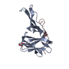
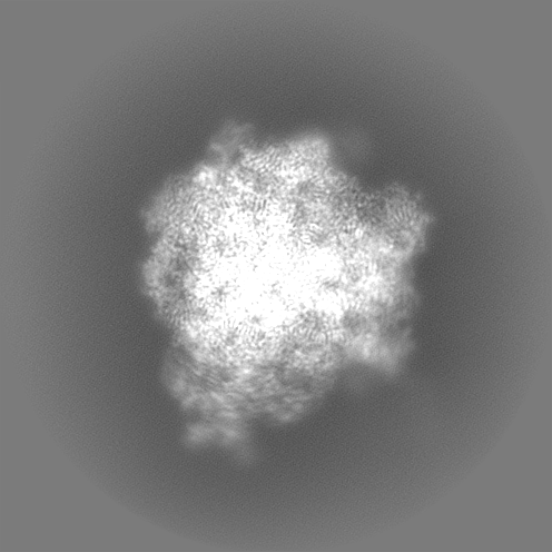
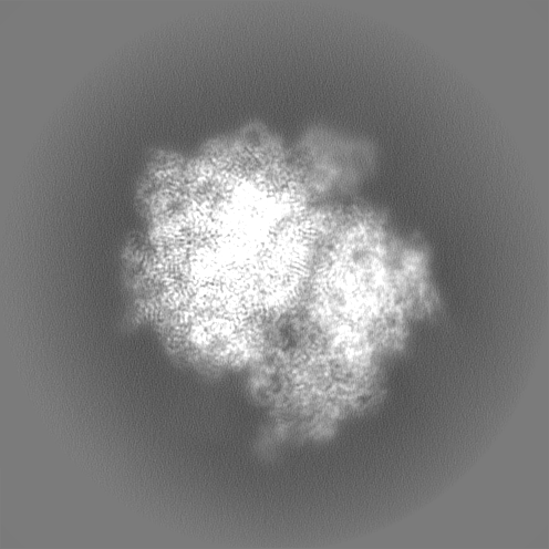
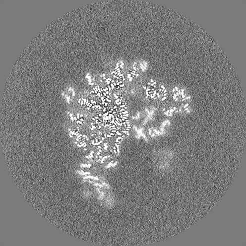
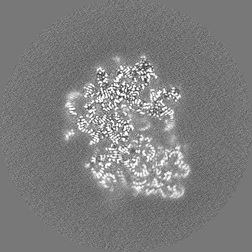
- EMDB-29788: Structure of WT E.coli ribosome 50S subunit with complexed with m... -
+
データを開く
IDまたはキーワード:
読み込み中...
-
基本情報
登録情報
データベース: EMDB / ID: EMD-29788
タイトル
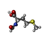
Structure of WT E.coli ribosome 50S subunit with complexed with mRNA, P-site fMet-NH-tRNAfMet and A-site 3-aminopyridine-4-carboxylic acid charged NH-tRNAPhe
negative regulation of cytoplasmic translational initiation / positive regulation of ribosome biogenesis / DnaA-L2 complex / negative regulation of DNA-templated DNA replication initiation / translational initiation / ribosome assembly / assembly of large subunit precursor of preribosome / cytosolic ribosome assembly / regulation of cell growth / ribosomal large subunit assembly ...negative regulation of cytoplasmic translational initiation / positive regulation of ribosome biogenesis / DnaA-L2 complex / negative regulation of DNA-templated DNA replication initiation / translational initiation / ribosome assembly / assembly of large subunit precursor of preribosome / cytosolic ribosome assembly / regulation of cell growth / ribosomal large subunit assembly / large ribosomal subunit / ribosome binding / transferase activity / 5S rRNA binding / large ribosomal subunit rRNA binding / cytosolic large ribosomal subunit / tRNA binding / cytoplasmic translation / rRNA binding / ribosome / structural constituent of ribosome / ribonucleoprotein complex / translation / response to antibiotic / RNA binding / zinc ion binding / cytoplasm / cytosol 類似検索 - 分子機能
Ribosomal protein L25, short-form / Ribosomal protein L31 type A / Ribosomal protein L31 signature. / Ribosomal protein L31 / Ribosomal protein L31 superfamily / Ribosomal protein L31 / Ribosomal protein L21, conserved site / Ribosomal protein L21 signature. / Ribosomal protein L16 signature 1. / : ...Ribosomal protein L25, short-form / Ribosomal protein L31 type A / Ribosomal protein L31 signature. / Ribosomal protein L31 / Ribosomal protein L31 superfamily / Ribosomal protein L31 / Ribosomal protein L21, conserved site / Ribosomal protein L21 signature. / Ribosomal protein L16 signature 1. / : / Ribosomal protein L6, conserved site / Ribosomal protein L6 signature 1. / Ribosomal protein L16, conserved site / Ribosomal protein L16 signature 2. / Ribosomal protein L9 signature. / Ribosomal protein L9, bacteria/chloroplast / Ribosomal protein L9, C-terminal / Ribosomal protein L9, C-terminal domain / Ribosomal protein L9, C-terminal domain superfamily / Ribosomal protein L17 signature. / Ribosomal L25p family / Ribosomal protein L25 / Ribosomal protein L36 signature. / Ribosomal protein L28/L24 superfamily / Ribosomal protein L25/Gln-tRNA synthetase, N-terminal / Ribosomal protein L25/Gln-tRNA synthetase, anti-codon-binding domain superfamily / Ribosomal protein L32p, bacterial type / Ribosomal protein L9, N-terminal domain superfamily / Ribosomal protein L9 / Ribosomal protein L9, N-terminal / Ribosomal protein L9, N-terminal domain / Ribosomal protein L28 / Ribosomal protein L35, conserved site / Ribosomal protein L35 signature. / Ribosomal protein L33, conserved site / Ribosomal protein L33 signature. / Ribosomal protein L35, non-mitochondrial / Ribosomal protein L5, bacterial-type / Ribosomal protein L18, bacterial-type / Ribosomal protein L6, bacterial-type / Ribosomal protein L19, conserved site / Ribosomal protein L19 signature. / Ribosomal protein L9/RNase H1, N-terminal / Ribosomal protein L36 / Ribosomal protein L36 superfamily / Ribosomal protein L36 / Ribosomal protein L20 signature. / Ribosomal protein L27, conserved site / Ribosomal protein L27 signature. / Ribosomal protein L14P, bacterial-type / Ribosomal protein L34, conserved site / Ribosomal protein L34 signature. / Ribosomal protein L22, bacterial/chloroplast-type / Ribosomal protein L2, bacterial/organellar-type / Ribosomal protein L35 / Ribosomal protein L35 superfamily / Ribosomal protein L35 / Ribosomal L28 family / Ribosomal protein L33 / Ribosomal protein L33 / Ribosomal protein L28/L24 / Ribosomal protein L18 / Ribosomal L18 of archaea, bacteria, mitoch. and chloroplast / Ribosomal protein L33 superfamily / Ribosomal protein L30, bacterial-type / : / Ribosomal protein L16 / L28p-like / Ribosomal protein L20 / Ribosomal protein L20 / Ribosomal protein L20, C-terminal / Ribosomal protein L21 / Ribosomal protein L27 / Ribosomal L27 protein / Ribosomal protein L19 / Ribosomal protein L19 superfamily / Ribosomal protein L19 / Ribosomal proteins 50S L24/mitochondrial 39S L24 / Ribosomal protein L17 / Ribosomal protein L17 superfamily / Ribosomal protein L17 / Ribosomal protein L21-like / L21-like superfamily / Ribosomal prokaryotic L21 protein / Ribosomal L32p protein family / Ribosomal protein L32p / Ribosomal protein L24 / Ribosomal protein L34 / Ribosomal protein L34 / Ribosomal protein L13, bacterial-type / Ribosomal protein L3, bacterial/organelle-type / Ribosomal protein L15, bacterial-type / 50S ribosomal protein uL4 / Ribosomal protein L23/L25, conserved site / Ribosomal protein L23 signature. / Ribosomal protein L30, conserved site / Ribosomal protein L30 signature. / Ribosomal protein L5, conserved site / Ribosomal protein L5 signature. / Ribosomal protein L2 signature. 類似検索 - ドメイン・相同性
Large ribosomal subunit protein uL15 / Large ribosomal subunit protein bL36 / Large ribosomal subunit protein bL28 / Large ribosomal subunit protein uL24 / Large ribosomal subunit protein bL17 / Large ribosomal subunit protein uL5 / Large ribosomal subunit protein uL23 / Large ribosomal subunit protein bL21 / 50S ribosomal protein L6 / 50S ribosomal protein L16 ...Large ribosomal subunit protein uL15 / Large ribosomal subunit protein bL36 / Large ribosomal subunit protein bL28 / Large ribosomal subunit protein uL24 / Large ribosomal subunit protein bL17 / Large ribosomal subunit protein uL5 / Large ribosomal subunit protein uL23 / Large ribosomal subunit protein bL21 / 50S ribosomal protein L6 / 50S ribosomal protein L16 / Large ribosomal subunit protein uL4 / Large ribosomal subunit protein bL20 / Large ribosomal subunit protein bL34 / Large ribosomal subunit protein uL29 / Large ribosomal subunit protein bL25 / Large ribosomal subunit protein bL32 / Large ribosomal subunit protein uL18 / Large ribosomal subunit protein bL33 / Large ribosomal subunit protein bL27 / Large ribosomal subunit protein bL31 / Large ribosomal subunit protein bL35 / Large ribosomal subunit protein uL2 / Large ribosomal subunit protein uL22 / Large ribosomal subunit protein uL13 / Large ribosomal subunit protein uL14 / Large ribosomal subunit protein uL3 / Large ribosomal subunit protein bL19 / Large ribosomal subunit protein uL30 / Large ribosomal subunit protein bL9 類似検索 - 構成要素
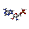
ジャーナル: ACS Cent Sci / 年: 2023 タイトル: Aminobenzoic Acid Derivatives Obstruct Induced Fit in the Catalytic Center of the Ribosome. 著者: Chandrima Majumdar / Joshua A Walker / Matthew B Francis / Alanna Schepartz / Jamie H D Cate / 要旨: The () ribosome can incorporate a variety of non-l-α-amino acid monomers into polypeptide chains but with poor efficiency. Although these monomers span a diverse set of compounds, there exists no ...The () ribosome can incorporate a variety of non-l-α-amino acid monomers into polypeptide chains but with poor efficiency. Although these monomers span a diverse set of compounds, there exists no high-resolution structural information regarding their positioning within the catalytic center of the ribosome, the peptidyl transferase center (PTC). Thus, details regarding the mechanism of amide bond formation and the structural basis for differences and defects in incorporation efficiency remain unknown. Within a set of three aminobenzoic acid derivatives-3-aminopyridine-4-carboxylic acid (Apy), aminobenzoic acid (ABZ), and aminobenzoic acid (ABZ)-the ribosome incorporates Apy into polypeptide chains with the highest efficiency, followed by ABZ and then ABZ, a trend that does not track with the nucleophilicity of the reactive amines. Here, we report high-resolution cryo-EM structures of the ribosome with each of these three aminobenzoic acid derivatives charged on tRNA bound in the aminoacyl-tRNA site (A-site). The structures reveal how the aromatic ring of each monomer sterically blocks the positioning of nucleotide U2506, thereby preventing rearrangement of nucleotide U2585 and the resulting induced fit in the PTC required for efficient amide bond formation. They also reveal disruptions to the bound water network that is believed to facilitate formation and breakdown of the tetrahedral intermediate. Together, the cryo-EM structures reported here provide a mechanistic rationale for differences in reactivity of aminobenzoic acid derivatives relative to l-α-amino acids and each other and identify stereochemical constraints on the size and geometry of non-monomers that can be accepted efficiently by wild-type ribosomes.
 ムービー
ムービー コントローラー


 データを開く
 基本情報
基本情報
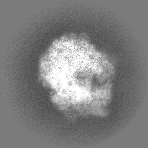
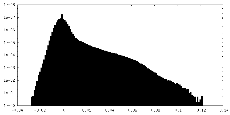
 マップデータ
 試料
試料 キーワード
キーワード 機能・相同性情報
 データ登録者
 米国, 1件
  引用
引用 構造の表示
構造の表示
 ダウンロードとリンク
 emd_29788.png
 http://ftp.pdbj.org/pub/emdb/structures/EMD-29788


 Z
Z Y
Y X
X


 試料の構成要素
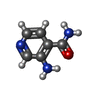

 解析
解析 電子顕微鏡法
 FIELD EMISSION GUN