 Movie
Movie Controller

[English] 日本語
 Yorodumi
- EMDB-2918: Outwardly curved tubulin sheet at the tip of a growing microtubule -
+ Open data

- Basic information
| Entry | Database: EMDB / ID: EMD-2918 | |||||||||
|---|---|---|---|---|---|---|---|---|---|---|
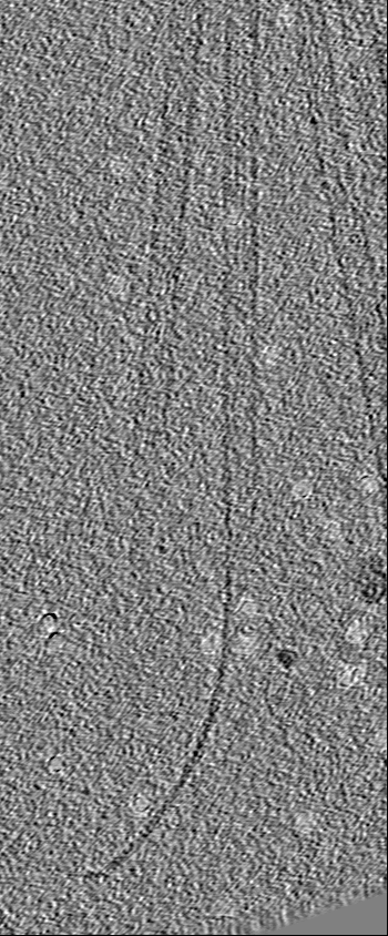
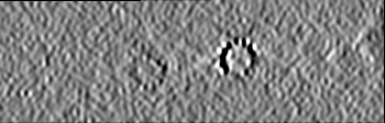
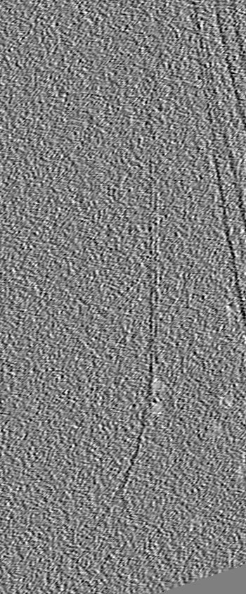
| Title | Outwardly curved tubulin sheet at the tip of a growing microtubule | |||||||||
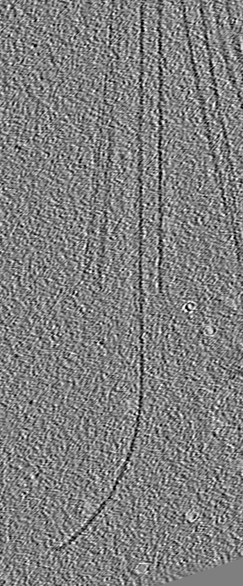
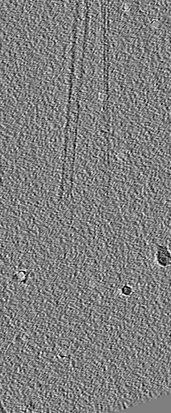
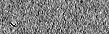
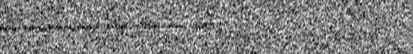
 Map data Map data | End of a microtubule assembled in the presence of EB1-gold nanoparticles (9.8 nm diameter). Nanoparticles were partially erased before 3D reconstruction. | |||||||||
 Sample Sample |
| |||||||||
 Keywords Keywords | End-binding one protein / tubulin / microtubule / functionalized gold nanoparticles / GTP-cap / GTP-hydrolysis | |||||||||
| Function / homology |  Function and homology information Function and homology informationprotein localization to astral microtubule / cortical microtubule cytoskeleton / mitotic spindle astral microtubule end / protein localization to microtubule / microtubule plus-end / cell projection membrane / attachment of mitotic spindle microtubules to kinetochore / microtubule plus-end binding / microtubule bundle formation / non-motile cilium assembly ...protein localization to astral microtubule / cortical microtubule cytoskeleton / mitotic spindle astral microtubule end / protein localization to microtubule / microtubule plus-end / cell projection membrane / attachment of mitotic spindle microtubules to kinetochore / microtubule plus-end binding / microtubule bundle formation / non-motile cilium assembly / protein localization to centrosome / microtubule organizing center / negative regulation of microtubule polymerization / mitotic spindle pole / cytoplasmic microtubule / establishment of mitotic spindle orientation / microtubule polymerization / microtubule-based process / spindle midzone / spindle assembly / regulation of microtubule polymerization or depolymerization / Amplification of signal from unattached kinetochores via a MAD2 inhibitory signal / Mitotic Prometaphase / EML4 and NUDC in mitotic spindle formation / Loss of Nlp from mitotic centrosomes / Loss of proteins required for interphase microtubule organization from the centrosome / Recruitment of mitotic centrosome proteins and complexes / Recruitment of NuMA to mitotic centrosomes / Anchoring of the basal body to the plasma membrane / positive regulation of microtubule polymerization / Resolution of Sister Chromatid Cohesion / AURKA Activation by TPX2 / ciliary basal body / RHO GTPases Activate Formins / Hydrolases; Acting on acid anhydrides; Acting on GTP to facilitate cellular and subcellular movement / structural constituent of cytoskeleton / microtubule cytoskeleton organization / Separation of Sister Chromatids / The role of GTSE1 in G2/M progression after G2 checkpoint / Regulation of PLK1 Activity at G2/M Transition / protein localization / cell migration / mitotic cell cycle / microtubule / hydrolase activity / cadherin binding / cell division / focal adhesion / GTPase activity / centrosome / GTP binding / protein kinase binding / Golgi apparatus / RNA binding / identical protein binding / metal ion binding / cytosol / cytoplasm Similarity search - Function | |||||||||
| Biological species |  Homo sapiens (human) / Homo sapiens (human) /  | |||||||||
| Method | electron tomography / cryo EM / negative staining | |||||||||
 Authors Authors | Guesdon A / Bazile F / Buey RM / Mohan R / Monier S / Angevin M / Heichette C / Tampe R / Duchesne L / Akhmanova A ...Guesdon A / Bazile F / Buey RM / Mohan R / Monier S / Angevin M / Heichette C / Tampe R / Duchesne L / Akhmanova A / Steinmetz MO / Chretien D | |||||||||
 Citation Citation | Journal: Nat Cell Biol / Year: 2016 Title: EB1 interacts with outwardly curved and straight regions of the microtubule lattice. Authors: Audrey Guesdon / Franck Bazile / Rubén M Buey / Renu Mohan / Solange Monier / Ruddi Rodríguez García / Morgane Angevin / Claire Heichette / Ralph Wieneke / Robert Tampé / Laurence ...Authors: Audrey Guesdon / Franck Bazile / Rubén M Buey / Renu Mohan / Solange Monier / Ruddi Rodríguez García / Morgane Angevin / Claire Heichette / Ralph Wieneke / Robert Tampé / Laurence Duchesne / Anna Akhmanova / Michel O Steinmetz / Denis Chrétien /      Abstract: EB1 is a microtubule plus-end tracking protein that recognizes GTP-tubulin dimers in microtubules and thus represents a unique probe to investigate the architecture of the GTP cap of growing ...EB1 is a microtubule plus-end tracking protein that recognizes GTP-tubulin dimers in microtubules and thus represents a unique probe to investigate the architecture of the GTP cap of growing microtubule ends. Here, we conjugated EB1 to gold nanoparticles (EB1-gold) and imaged by cryo-electron tomography its interaction with dynamic microtubules assembled in vitro from purified tubulin. EB1-gold forms comets at the ends of microtubules assembled in the presence of GTP, and interacts with the outer surface of curved and straight tubulin sheets as well as closed regions of the microtubule lattice. Microtubules assembled in the presence of GTP, different GTP analogues or cell extracts display similarly curved sheets at their growing ends, which gradually straighten as their protofilament number increases until they close into a tube. Together, our data provide unique structural information on the interaction of EB1 with growing microtubule ends. They further offer insights into the conformational changes that tubulin dimers undergo during microtubule assembly and the architecture of the GTP-cap region. | |||||||||
| History |
|
- Structure visualization
| Movie |
Movie viewer |
|---|---|
| Structure viewer | EM map: SurfViewMolmilJmol/JSmol |
| Supplemental images |
 UCSF Chimera
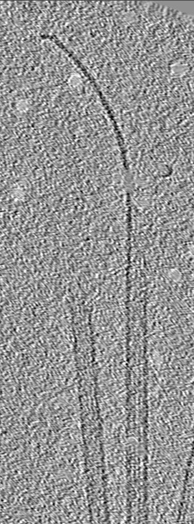
- Downloads & links
-EMDB archive
| Map data | emd_2918.map.gz | 48.9 MB | EMDB map data format | |
|---|---|---|---|---|
| Header (meta data) | emd-2918-v30.xmlemd-2918.xml | 14.2 KB 14.2 KB | Display Display | EMDB header |
| Images | emd_2918.tif | 211.3 KB | ||
| Archive directory |  http://ftp.pdbj.org/pub/emdb/structures/EMD-2918ftp://ftp.pdbj.org/pub/emdb/structures/EMD-2918 http://ftp.pdbj.org/pub/emdb/structures/EMD-2918ftp://ftp.pdbj.org/pub/emdb/structures/EMD-2918 | HTTPS FTP |
-Validation report
| Summary document | emd_2918_validation.pdf.gz | 169.4 KB | Display | EMDB validaton report |
|---|---|---|---|---|
| Full document | emd_2918_full_validation.pdf.gz | 168.5 KB | Display | |
| Data in XML | emd_2918_validation.xml.gz | 3.6 KB | Display | |
| Arichive directory | https://ftp.pdbj.org/pub/emdb/validation_reports/EMD-2918ftp://ftp.pdbj.org/pub/emdb/validation_reports/EMD-2918 | HTTPS FTP |
-Related structure data
| Related structure data |  2912C  2915C  2916C  2919C  2920C  2921C C: citing same article ( |
|---|---|
| Similar structure data |
-Links
| EMDB pages | EMDB (EBI/PDBe) / EMDataResource |
|---|---|
| Related items in Molecule of the Month |


-Map
| File | Download / File: emd_2918.map.gz / Format: CCP4 / Size: 61.6 MB / Type: IMAGE STORED AS SIGNED INTEGER (2 BYTES) | ||||||||||||||||||||||||||||||||||||||||||||||||||||||||||||||||||||
|---|---|---|---|---|---|---|---|---|---|---|---|---|---|---|---|---|---|---|---|---|---|---|---|---|---|---|---|---|---|---|---|---|---|---|---|---|---|---|---|---|---|---|---|---|---|---|---|---|---|---|---|---|---|---|---|---|---|---|---|---|---|---|---|---|---|---|---|---|---|
| Annotation | End of a microtubule assembled in the presence of EB1-gold nanoparticles (9.8 nm diameter). Nanoparticles were partially erased before 3D reconstruction. | ||||||||||||||||||||||||||||||||||||||||||||||||||||||||||||||||||||
| Projections & slices | Image control
Images are generated by Spider. generated in cubic-lattice coordinate | ||||||||||||||||||||||||||||||||||||||||||||||||||||||||||||||||||||
| Voxel size | X=Y=Z: 8.7 Å | ||||||||||||||||||||||||||||||||||||||||||||||||||||||||||||||||||||
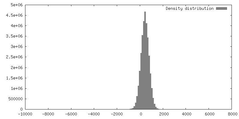
| Density |
| ||||||||||||||||||||||||||||||||||||||||||||||||||||||||||||||||||||
| Symmetry | Space group: 1 | ||||||||||||||||||||||||||||||||||||||||||||||||||||||||||||||||||||
| Details | EMDB XML:
CCP4 map header:
| ||||||||||||||||||||||||||||||||||||||||||||||||||||||||||||||||||||
 Z (Sec.)
 Y (Row.)
 X (Col.)


-Supplemental data
- Sample components
-Entire : EB1 conjugated to 9.8 nm gold nanoparticles, in interaction with ...
| Entire | Name: EB1 conjugated to 9.8 nm gold nanoparticles, in interaction with a microtubule growing end. |
|---|---|
| Components |
|
-Supramolecule #1000: EB1 conjugated to 9.8 nm gold nanoparticles, in interaction with ...
| Supramolecule | Name: EB1 conjugated to 9.8 nm gold nanoparticles, in interaction with a microtubule growing end. type: sample / ID: 1000 / Details: The sample was monodisperse Oligomeric state: EB1: homodimer. Microtubule: polymer of tubulin. Number unique components: 3 |
|---|
-Macromolecule #1: End-binding protein one
| Macromolecule | Name: End-binding protein one / type: protein_or_peptide / ID: 1 / Name.synonym: EB1 Details: EB1 with a haxa-histidine tag inserted into its C-terminal extremity was conjugated to 9.8 nanometer gold nanoparticles functionalized with Ni-trisNTA groups Oligomeric state: Homodimer / Recombinant expression: Yes |
|---|---|
| Source (natural) | Organism: Homo sapiens (human) / synonym: Human / Location in cell: Cytoplasm |
| Molecular weight | Experimental: 30 KDa / Theoretical: 30 KDa |
| Recombinant expression | Organism:  |
| Sequence | UniProtKB: Microtubule-associated protein RP/EB family member 1 |
-Macromolecule #2: Tubulin alpha chain
| Macromolecule | Name: Tubulin alpha chain / type: protein_or_peptide / ID: 2 Details: Heterodimer alpha-beta polymerized into microtubules Recombinant expression: No / Database: NCBI |
|---|---|
| Source (natural) | Organism: |
| Molecular weight | Theoretical: 500.68 KDa |
| Sequence | UniProtKB: Tubulin alpha-1A chain / GO: microtubule-based process / InterPro: Tubulin |
-Macromolecule #3: Tubulin beta chain
| Macromolecule | Name: Tubulin beta chain / type: protein_or_peptide / ID: 3 Details: Heterodimer alpha-beta polymerized into microtubules Recombinant expression: No / Database: NCBI |
|---|---|
| Source (natural) | Organism: |
| Molecular weight | Theoretical: 498.61 KDa |
| Sequence | UniProtKB: Tubulin beta chain / GO: microtubule-based process / InterPro: Tubulin, conserved site |
-Experimental details
-Structure determination
| Method | negative staining, cryo EM |
|---|---|
 Processing Processing | electron tomography |
| Aggregation state | particle |
-Sample preparation
| Buffer | pH: 6.8 Details: 80 mM Pipes, 1 mM MgCl2, 62.5 mM KCl, 1mM EGTA, 1 mM GTP |
|---|---|
| Staining | Type: NEGATIVE / Details: Vitrified specimen. |
| Grid | Details: 300 mesh grid coated with home-made holey-carbon film. |
| Vitrification | Cryogen name: ETHANE / Chamber humidity: 90 % / Chamber temperature: 93 K / Instrument: HOMEMADE PLUNGER Details: Specimen maintained at 35 degrees celcius in saturated humidity conditions before vitrification. Timed resolved state: Specimen frozen ~3 min after the beginning of assembly. Method: Assembly in a test tube at 35 degrees celcius for ~3 min, deposit of a 4 microliter onto the grid, blotting for ~2 seconds and rapid plunging into liquid ethane. |
- Electron microscopy
| Microscope | FEI TECNAI 20 |
|---|---|
| Temperature | Average: 174 K |
| Alignment procedure | Legacy - Astigmatism: Astigmatism corrected at high magnification |
| Details | Tilt serie started from zero. Saxton acquisition scheme with 1.9 degrees increments. 79 images acquired in post-tracking mode. Tilt series between -63.3 and 55.9 degrees after correction.Camera used in binning mode 2, 0.87 nm pixel size. |
| Date | Oct 18, 2013 |
| Image recording | Category: CCD / Film or detector model: GATAN ULTRASCAN 1000 (2k x 2k) / Number real images: 79 / Average electron dose: 0.3 e/Å2 Details: Camera used in binning mode 2, at a final pixel size of 0.87 nm. Bits/pixel: 16 |
| Electron beam | Acceleration voltage: 200 kV / Electron source: LAB6 |
| Electron optics | Illumination mode: FLOOD BEAM / Imaging mode: BRIGHT FIELD / Cs: 2.0 mm / Nominal defocus max: 1.5 µm / Nominal defocus min: 1.5 µm / Nominal magnification: 25000 |
| Sample stage | Specimen holder: Liquid nitrogen cooled / Specimen holder model: GATAN LIQUID NITROGEN / Tilt series - Axis1 - Min angle: -60 ° / Tilt series - Axis1 - Max angle: 60 ° / Tilt series - Axis1 - Angle increment: 2 ° |
-Image processing
| Details | 3D reconstruction performed using eTomo from IMOD software. Reconstruction by backprojection, using a Radial filter cutoffof 0.15 and a faloff of 0.05. Nanoparticles partially erased before 3D reconstruction. |
|---|---|
| Final reconstruction | Algorithm: OTHER / Software - Name: eTomo, IMOD Details: Backprojection using a radial filter cutoff of 0.15 and a faloff of 0.05. Number images used: 79 |
