登録情報 データベース : EMDB / ID : EMD-28741タイトル Brain-derived 42-residue amyloid-beta fibril type B 複合体 : amyloid-b 42 (Ab42) fibrilタンパク質・ペプチド : Beta-amyloid protein 42 / / / / 機能・相同性 分子機能 ドメイン・相同性 構成要素
/ / / / / / / / / / / / / / / / / / / / / / / / / / / / / / / / / / / / / / / / / / / / / / / / / / / / / / / / / / / / / / / / / / / / / / / / / / / / / / / / / / / / / / / / / / / / / / / / / / / / / / / / / / / / / / / / / / / / / / / / / / / / / / / / / / / / / / / / / / / / / / / / / / / 生物種 Homo sapiens (ヒト)手法 / / 解像度 : 2.76 Å Tycko R / Lee M / Yau Y-M / Louis JM 資金援助 Organization Grant number 国 Not funded
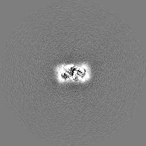
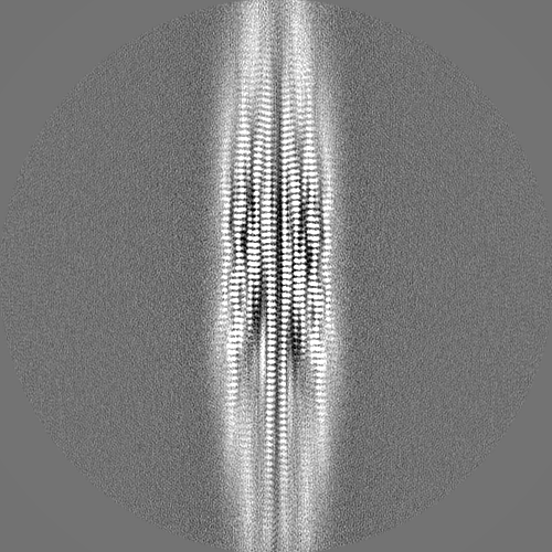
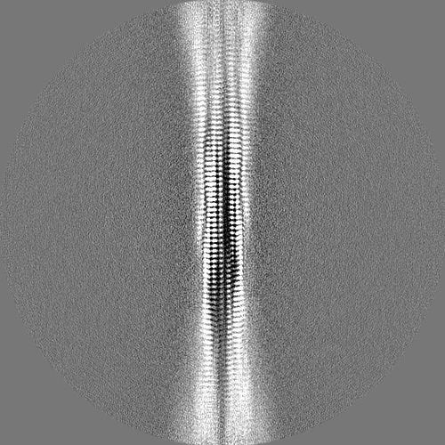
ジャーナル : Proc Natl Acad Sci U S A / 年 : 2023タイトル : Structures of brain-derived 42-residue amyloid-β fibril polymorphs with unusual molecular conformations and intermolecular interactions.著者 : Myungwoon Lee / Wai-Ming Yau / John M Louis / Robert Tycko / 要旨 : Fibrils formed by the 42-residue amyloid-β peptide (Aβ42), a main component of amyloid deposits in Alzheimer's disease (AD), are known to be polymorphic, i.e., to contain multiple possible ... Fibrils formed by the 42-residue amyloid-β peptide (Aβ42), a main component of amyloid deposits in Alzheimer's disease (AD), are known to be polymorphic, i.e., to contain multiple possible molecular structures. Previous studies of Aβ42 fibrils, including fibrils prepared entirely in vitro or extracted from brain tissue and using solid-state NMR (ssNMR) or cryogenic electron microscopy (cryo-EM) methods, have found polymorphs with differences in amino acid sidechain orientations, lengths of structurally ordered segments, and contacts between cross-β subunit pairs within a single filament. Despite these differences, Aβ42 molecules adopt a common S-shaped conformation in all previously described high-resolution Aβ42 fibril structures. Here we report two cryo-EM-based structures of Aβ42 fibrils that are qualitatively different, in samples derived from AD brain tissue by seeded growth. In type A fibrils, residues 12 to 42 adopt a ν-shaped conformation, with both intra-subunit and intersubunit hydrophobic contacts to form a compact core. In type B fibrils, residues 2 to 42 adopt an υ-shaped conformation, with only intersubunit contacts and internal pores. Type A and type B fibrils have opposite helical handedness. Cryo-EM density maps and molecular dynamics simulations indicate intersubunit K16-A42 salt bridges in type B fibrils and partially occupied K28-A42 salt bridges in type A fibrils. The coexistence of two predominant polymorphs, with differences in N-terminal dynamics, is supported by ssNMR data, as is faithful propagation of structures from first-generation to second-generation brain-seeded Aβ42 fibril samples. These results demonstrate that Aβ42 fibrils can exhibit a greater range of structural variations than seen in previous studies. 履歴 登録 2022年10月31日 - ヘッダ(付随情報) 公開 2023年3月22日 - マップ公開 2023年3月22日 - 更新 2024年6月19日 - 現状 2024年6月19日 処理サイト : RCSB / 状態 : 公開
すべて表示 表示を減らす
 ムービー
ムービー コントローラー


 データを開く
 基本情報
基本情報
 マップデータ
 試料
試料 キーワード
キーワード 機能・相同性情報
 Homo sapiens (ヒト)
 データ登録者
 米国, 1件
  引用
引用 構造の表示
構造の表示
 ダウンロードとリンク
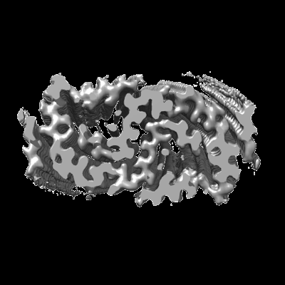 emd_28741.png
 http://ftp.pdbj.org/pub/emdb/structures/EMD-28741


 Z
Z Y
Y X
X


 試料の構成要素
 解析
解析 電子顕微鏡法
 FIELD EMISSION GUN