+ Open data
- Basic information
| Entry |  | |||||||||
|---|---|---|---|---|---|---|---|---|---|---|
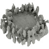
| Title | Subtomogram of Fluad vaccine hemagglutinin spiked nanodisc | |||||||||
 Map data Map data | subtomogram of Fluad nanodisc, raw | |||||||||
 Sample Sample |
| |||||||||
| Biological species |   Influenza A virus Influenza A virus | |||||||||
| Method | electron tomography / cryo EM | |||||||||
 Authors Authors | Gallagher JR / Audray AK | |||||||||
| Funding support |  United States, 1 items United States, 1 items
| |||||||||
 Citation Citation |  Journal: Nat Commun / Year: 2023 Journal: Nat Commun / Year: 2023Title: Commercial influenza vaccines vary in HA-complex structure and in induction of cross-reactive HA antibodies. Authors: Mallory L Myers / John R Gallagher / Alexander J Kim / Walker H Payne / Samantha Maldonado-Puga / Haralabos Assimakopoulos / Kevin W Bock / Udana Torian / Ian N Moore / Audray K Harris /  Abstract: Influenza virus infects millions of people annually and can cause global pandemics. Hemagglutinin (HA) is the primary component of commercial influenza vaccines (CIV), and antibody titer to HA is a ...Influenza virus infects millions of people annually and can cause global pandemics. Hemagglutinin (HA) is the primary component of commercial influenza vaccines (CIV), and antibody titer to HA is a primary correlate of protection. Continual antigenic variation of HA requires that CIVs are reformulated yearly. Structural organization of HA complexes have not previously been correlated with induction of broadly reactive antibodies, yet CIV formulations vary in how HA is organized. Using electron microscopy to study four current CIVs, we find structures including: individual HAs, starfish structures with up to 12 HA molecules, and novel spiked-nanodisc structures that display over 50 HA molecules along the complex's perimeter. CIV containing these spiked nanodiscs elicit the highest levels of heterosubtypic cross-reactive antibodies in female mice. Here, we report that HA structural organization can be an important CIV parameter and can be associated with the induction of cross-reactive antibodies to conserved HA epitopes. | |||||||||
| History |
|
- Structure visualization
| Supplemental images |
|---|
- Downloads & links
-EMDB archive
| Map data |  emd_27232.map.gz emd_27232.map.gz | 14.7 MB |  EMDB map data format EMDB map data format | |
|---|---|---|---|---|
| Header (meta data) |  emd-27232-v30.xml emd-27232-v30.xml emd-27232.xml emd-27232.xml | 12.3 KB 12.3 KB | Display Display |  EMDB header EMDB header |
| Images |  emd_27232.png emd_27232.png | 163.8 KB | ||
| Others |  emd_27232_additional_1.map.gz emd_27232_additional_1.map.gz | 2.6 MB | ||
| Archive directory |  http://ftp.pdbj.org/pub/emdb/structures/EMD-27232 http://ftp.pdbj.org/pub/emdb/structures/EMD-27232 ftp://ftp.pdbj.org/pub/emdb/structures/EMD-27232 ftp://ftp.pdbj.org/pub/emdb/structures/EMD-27232 | HTTPS FTP |
-Validation report
| Summary document |  emd_27232_validation.pdf.gz emd_27232_validation.pdf.gz | 797.1 KB | Display |  EMDB validaton report EMDB validaton report |
|---|---|---|---|---|
| Full document |  emd_27232_full_validation.pdf.gz emd_27232_full_validation.pdf.gz | 796.6 KB | Display | |
| Data in XML |  emd_27232_validation.xml.gz emd_27232_validation.xml.gz | 3 KB | Display | |
| Data in CIF |  emd_27232_validation.cif.gz emd_27232_validation.cif.gz | 3.5 KB | Display | |
| Arichive directory |  https://ftp.pdbj.org/pub/emdb/validation_reports/EMD-27232 https://ftp.pdbj.org/pub/emdb/validation_reports/EMD-27232 ftp://ftp.pdbj.org/pub/emdb/validation_reports/EMD-27232 ftp://ftp.pdbj.org/pub/emdb/validation_reports/EMD-27232 | HTTPS FTP |
-Related structure data
- Links
Links
| EMDB pages |  EMDB (EBI/PDBe) / EMDB (EBI/PDBe) /  EMDataResource EMDataResource |
|---|
- Map
Map
| File |  Download / File: emd_27232.map.gz / Format: CCP4 / Size: 25.1 MB / Type: IMAGE STORED AS SIGNED BYTE Download / File: emd_27232.map.gz / Format: CCP4 / Size: 25.1 MB / Type: IMAGE STORED AS SIGNED BYTE | ||||||||||||||||||||
|---|---|---|---|---|---|---|---|---|---|---|---|---|---|---|---|---|---|---|---|---|---|
| Annotation | subtomogram of Fluad nanodisc, raw | ||||||||||||||||||||
| Voxel size | X=Y=Z: 4.6057 Å | ||||||||||||||||||||
| Density |
| ||||||||||||||||||||
| Symmetry | Space group: 1 | ||||||||||||||||||||
| Details | EMDB XML:
|
-Supplemental data
-Additional map: subtomogram of Fluad nanodisc, processed with nonlinear anisotropic...
| File | emd_27232_additional_1.map | ||||||||||||
|---|---|---|---|---|---|---|---|---|---|---|---|---|---|
| Annotation | subtomogram of Fluad nanodisc, processed with nonlinear anisotropic diffusion. | ||||||||||||
| Projections & Slices |
| ||||||||||||
| Density Histograms |
- Sample components
-Entire : Hemagglutinin protein extracted from these influenza viruses: A/S...
| Entire | Name: Hemagglutinin protein extracted from these influenza viruses: A/Singapore/GP1908/2015 A/Singapore/INFIMH-16-0019/2016 B/Maryland/15/2016 |
|---|---|
| Components |
|
-Supramolecule #1: Hemagglutinin protein extracted from these influenza viruses: A/S...
| Supramolecule | Name: Hemagglutinin protein extracted from these influenza viruses: A/Singapore/GP1908/2015 A/Singapore/INFIMH-16-0019/2016 B/Maryland/15/2016 type: complex / ID: 1 / Chimera: Yes / Parent: 0 / Details: formulated in MF59C.1 adjuvant |
|---|---|
| Source (natural) | Organism:   Influenza A virus Influenza A virus |
-Supramolecule #2: A/Singapore/INFIMH-16-0019/2016 hemagglutinin protein
| Supramolecule | Name: A/Singapore/INFIMH-16-0019/2016 hemagglutinin protein / type: complex / ID: 2 / Chimera: Yes / Parent: 1 / Details: formulated in MF59C.1 adjuvant |
|---|
-Supramolecule #3: A/Singapore/GP1908/2015 hemagglutinin protein
| Supramolecule | Name: A/Singapore/GP1908/2015 hemagglutinin protein / type: complex / ID: 3 / Chimera: Yes / Parent: 1 / Details: formulated with MF59C.1 adjuvant |
|---|
-Supramolecule #4: B/Maryland/15/2016 hemagglutinin protein
| Supramolecule | Name: B/Maryland/15/2016 hemagglutinin protein / type: complex / ID: 4 / Chimera: Yes / Parent: 1 / Details: formulated with MF59C.1 adjuvant |
|---|
-Experimental details
-Structure determination
| Method | cryo EM |
|---|---|
 Processing Processing | electron tomography |
| Aggregation state | particle |
- Sample preparation
| Buffer | pH: 7.4 |
|---|---|
| Grid | Model: C-flat-2/2 / Material: COPPER / Mesh: 200 / Support film - Material: CARBON / Support film - topology: HOLEY |
| Vitrification | Cryogen name: ETHANE / Chamber humidity: 100 % / Chamber temperature: 310 K / Instrument: LEICA EM GP |
| Sectioning | Other: NO SECTIONING |
| Fiducial marker | Manufacturer: Electron Microscopy Sciences / Diameter: 10 nm |
- Electron microscopy
| Microscope | FEI TITAN KRIOS |
|---|---|
| Specialist optics | Phase plate: VOLTA PHASE PLATE / Energy filter - Slit width: 20 eV |
| Image recording | Film or detector model: FEI FALCON III (4k x 4k) / Average electron dose: 1.64 e/Å2 |
| Electron beam | Acceleration voltage: 300 kV / Electron source:  FIELD EMISSION GUN FIELD EMISSION GUN |
| Electron optics | Illumination mode: FLOOD BEAM / Imaging mode: BRIGHT FIELD / Nominal defocus max: 0.5 µm / Nominal defocus min: 0.2 µm |
| Experimental equipment |  Model: Titan Krios / Image courtesy: FEI Company |
- Image processing
| Final reconstruction | Software - Name:  IMOD / Number images used: 61 IMOD / Number images used: 61 |
|---|
 Movie
Movie Controller


 Z
Z Y
Y X
X


