 ムービー
ムービー コントローラー


+ データを開く

- 基本情報
基本情報
| 登録情報 |  | |||||||||
|---|---|---|---|---|---|---|---|---|---|---|
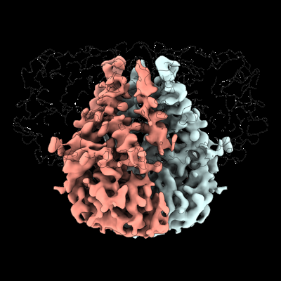
| タイトル | Cryo-EM structure of SARS-CoV-2 M protein in a lipid nanodisc | |||||||||
 マップデータ マップデータ | ||||||||||
 試料 試料 |
| |||||||||
 キーワード キーワード | SARS-COV-2 / CORONAVIRUS / VIRAL PROTEIN / CAPSID PROTEIN / MEMBRANE PROTEIN | |||||||||
| 機能・相同性 |  機能・相同性情報 機能・相同性情報Maturation of protein M / SARS-CoV-2 modulates autophagy / cytoplasmic capsid assembly / host cell Golgi membrane / CD28 dependent PI3K/Akt signaling / endoplasmic reticulum-Golgi intermediate compartment / SARS-CoV-2 targets host intracellular signalling and regulatory pathways / protein sequestering activity / symbiont-mediated suppression of host cytoplasmic pattern recognition receptor signaling pathway via inhibition of MAVS activity / VEGFR2 mediated vascular permeability ...Maturation of protein M / SARS-CoV-2 modulates autophagy / cytoplasmic capsid assembly / host cell Golgi membrane / CD28 dependent PI3K/Akt signaling / endoplasmic reticulum-Golgi intermediate compartment / SARS-CoV-2 targets host intracellular signalling and regulatory pathways / protein sequestering activity / symbiont-mediated suppression of host cytoplasmic pattern recognition receptor signaling pathway via inhibition of MAVS activity / VEGFR2 mediated vascular permeability / PIP3 activates AKT signaling / TRAF3-dependent IRF activation pathway / Translation of Structural Proteins / Virion Assembly and Release / Induction of Cell-Cell Fusion / structural constituent of virion / Attachment and Entry / virus-mediated perturbation of host defense response / viral envelope / SARS-CoV-2 activates/modulates innate and adaptive immune responses / virion membrane / identical protein binding / plasma membrane 類似検索 - 分子機能 | |||||||||
| 生物種 |   Severe acute respiratory syndrome coronavirus 2 (ウイルス) Severe acute respiratory syndrome coronavirus 2 (ウイルス) | |||||||||
| 手法 | 単粒子再構成法 / クライオ電子顕微鏡法 / 解像度: 3.52 Å | |||||||||
 データ登録者 データ登録者 | Dolan KA / Brohawn SG | |||||||||
| 資金援助 |  米国, 1件 米国, 1件
| |||||||||
 引用 引用 | ジャーナル: Elife / 年: 2022 タイトル: Structure of SARS-CoV-2 M protein in lipid nanodiscs. 著者: Kimberly A Dolan / Mandira Dutta / David M Kern / Abhay Kotecha / Gregory A Voth / Stephen G Brohawn /  要旨: SARS-CoV-2 encodes four structural proteins incorporated into virions, spike (S), envelope (E), nucleocapsid (N), and membrane (M). M plays an essential role in viral assembly by organizing other ...SARS-CoV-2 encodes four structural proteins incorporated into virions, spike (S), envelope (E), nucleocapsid (N), and membrane (M). M plays an essential role in viral assembly by organizing other structural proteins through physical interactions and directing them to sites of viral budding. As the most abundant protein in the viral envelope and a target of patient antibodies, M is a compelling target for vaccines and therapeutics. Still, the structure of M and molecular basis for its role in virion formation are unknown. Here, we present the cryo-EM structure of SARS-CoV-2 M in lipid nanodiscs to 3.5 Å resolution. M forms a 50 kDa homodimer that is structurally related to the SARS-CoV-2 ORF3a viroporin, suggesting a shared ancestral origin. Structural comparisons reveal how intersubunit gaps create a small, enclosed pocket in M and large open cavity in ORF3a, consistent with a structural role and ion channel activity, respectively. M displays a strikingly electropositive cytosolic surface that may be important for interactions with N, S, and viral RNA. Molecular dynamics simulations show a high degree of structural rigidity in a simple lipid bilayer and support a role for M homodimers in scaffolding viral assembly. Together, these results provide insight into roles for M in coronavirus assembly and structure. #1: ジャーナル: Acta Crystallogr., Sect. D: Biol. Crystallogr.年: 2018 タイトル: Real-space refinement in PHENIX for cryo-EM and crystallography 著者: Afonine PV | |||||||||
| 履歴 |
|
- 構造の表示
構造の表示
| 添付画像 |
|---|
- ダウンロードとリンク
-EMDBアーカイブ
| マップデータ | emd_26993.map.gz | 85.8 MB | EMDBマップデータ形式 | |
|---|---|---|---|---|
| ヘッダ (付随情報) | emd-26993-v30.xmlemd-26993.xml | 15.9 KB 15.9 KB | 表示 表示 | EMDBヘッダ |
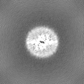
| 画像 |  emd_26993.png emd_26993.png | 116.7 KB | ||
| Filedesc metadata | emd-26993.cif.gz | 5.5 KB | ||
| その他 | emd_26993_half_map_1.map.gzemd_26993_half_map_2.map.gz | 84.5 MB 84.5 MB | ||
| アーカイブディレクトリ |  http://ftp.pdbj.org/pub/emdb/structures/EMD-26993ftp://ftp.pdbj.org/pub/emdb/structures/EMD-26993 http://ftp.pdbj.org/pub/emdb/structures/EMD-26993ftp://ftp.pdbj.org/pub/emdb/structures/EMD-26993 | HTTPS FTP |
-検証レポート
| 文書・要旨 | emd_26993_validation.pdf.gz | 911.9 KB | 表示 | EMDB検証レポート |
|---|---|---|---|---|
| 文書・詳細版 | emd_26993_full_validation.pdf.gz | 911.5 KB | 表示 | |
| XML形式データ | emd_26993_validation.xml.gz | 12.9 KB | 表示 | |
| CIF形式データ | emd_26993_validation.cif.gz | 15.3 KB | 表示 | |
| アーカイブディレクトリ | https://ftp.pdbj.org/pub/emdb/validation_reports/EMD-26993ftp://ftp.pdbj.org/pub/emdb/validation_reports/EMD-26993 | HTTPS FTP |
-関連構造データ
| 関連構造データ |  8ctkMC M: このマップから作成された原子モデル C: 同じ文献を引用 ( |
|---|---|
| 類似構造データ | |
| 電子顕微鏡画像生データ | EMPIAR-11067 (タイトル: Structure of SARS-CoV-2 M protein in lipid nanodiscs Data size: 4.0 TB Data #1: SARS-CoV-2 M protein in an MSP1E3D1 lipid nanodisc [micrographs - multiframe]) |
-リンク
| EMDBのページ | EMDB (EBI/PDBe) / EMDataResource |
|---|---|
| 「今月の分子」の関連する項目 |


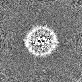
-マップ
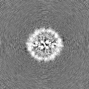
| ファイル | ダウンロード / ファイル: emd_26993.map.gz / 形式: CCP4 / 大きさ: 91.1 MB / タイプ: IMAGE STORED AS FLOATING POINT NUMBER (4 BYTES) | ||||||||||||||||||||
|---|---|---|---|---|---|---|---|---|---|---|---|---|---|---|---|---|---|---|---|---|---|
| ボクセルのサイズ | X=Y=Z: 0.727 Å | ||||||||||||||||||||
| 密度 |
| ||||||||||||||||||||
| 対称性 | 空間群: 1 | ||||||||||||||||||||
| 詳細 | EMDB XML:
|
-添付データ
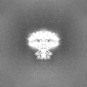
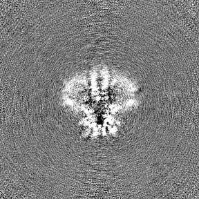
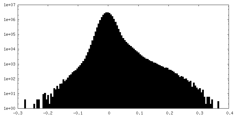
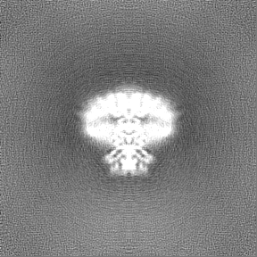
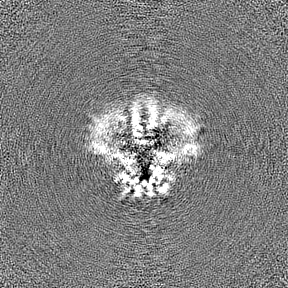
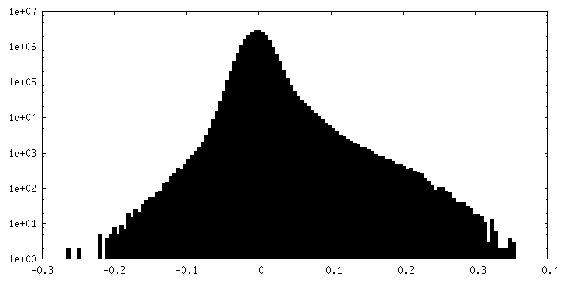
-ハーフマップ: #2
| ファイル | emd_26993_half_map_1.map | ||||||||||||
|---|---|---|---|---|---|---|---|---|---|---|---|---|---|
| 投影像・断面図 |
| ||||||||||||
| 密度ヒストグラム |
 Z
Z Y
Y X
X
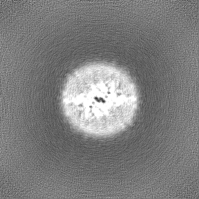


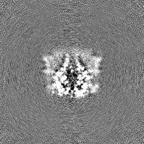
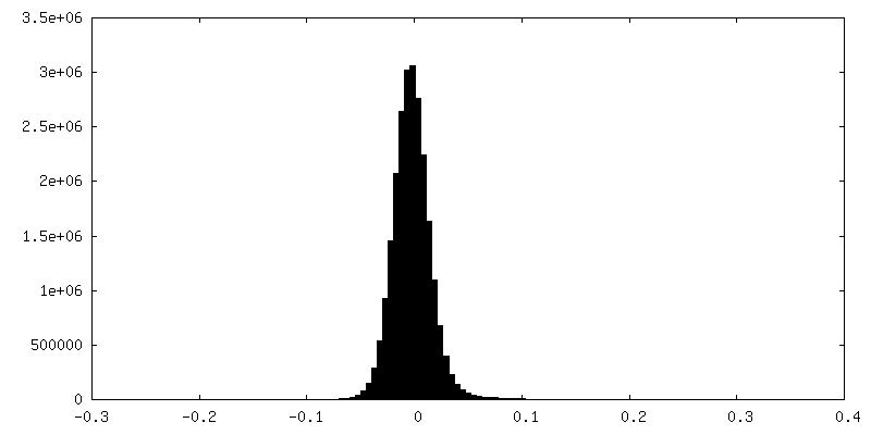
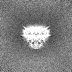
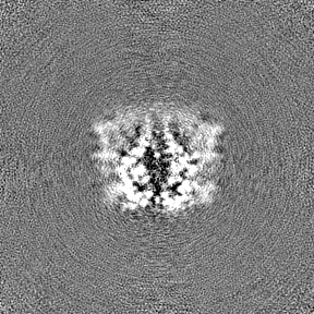
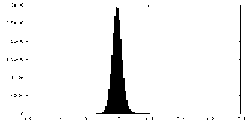
-ハーフマップ: #1
| ファイル | emd_26993_half_map_2.map | ||||||||||||
|---|---|---|---|---|---|---|---|---|---|---|---|---|---|
| 投影像・断面図 |
| ||||||||||||
| 密度ヒストグラム |
- 試料の構成要素
-全体 : M protein
| 全体 | 名称: M protein |
|---|---|
| 要素 |
|
-超分子 #1: M protein
| 超分子 | 名称: M protein / タイプ: complex / ID: 1 / 親要素: 0 / 含まれる分子: all |
|---|---|
| 由来(天然) | 生物種: Severe acute respiratory syndrome coronavirus 2 (ウイルス) |
| 分子量 | 理論値: 52 KDa |
-分子 #1: Membrane protein
| 分子 | 名称: Membrane protein / タイプ: protein_or_peptide / ID: 1 / コピー数: 2 / 光学異性体: LEVO |
|---|---|
| 由来(天然) | 生物種: Severe acute respiratory syndrome coronavirus 2 (ウイルス) |
| 分子量 | 理論値: 26.190693 KDa |
| 組換発現 | 生物種:   Spodoptera frugiperda (ツマジロクサヨトウ) Spodoptera frugiperda (ツマジロクサヨトウ) |
| 配列 | 文字列: MADSNGTITV EELKKLLEQW NLVIGFLFLT WICLLQFAYA NRNRFLYIIK LIFLWLLWPV TLACFVLAAV YRINWITGGI AIAMACLVG LMWLSYFIAS FRLFARTRSM WSFNPETNIL LNVPLHGTIL TRPLLESELV IGAVILRGHL RIAGHHLGRC D IKDLPKEI ...文字列: MADSNGTITV EELKKLLEQW NLVIGFLFLT WICLLQFAYA NRNRFLYIIK LIFLWLLWPV TLACFVLAAV YRINWITGGI AIAMACLVG LMWLSYFIAS FRLFARTRSM WSFNPETNIL LNVPLHGTIL TRPLLESELV IGAVILRGHL RIAGHHLGRC D IKDLPKEI TVATSRTLSY YKLGASQRVA GDSGFAAYSR YRIGNYKLNT DHSSSSDNIA LLVQSNSLEV LFQ UniProtKB: Membrane protein |
-実験情報
-構造解析
| 手法 | クライオ電子顕微鏡法 |
|---|---|
 解析 解析 | 単粒子再構成法 |
| 試料の集合状態 | particle |
-試料調製
| 濃度 | 1.3 mg/mL | |||||||||
|---|---|---|---|---|---|---|---|---|---|---|
| 緩衝液 | pH: 7.4 構成要素:
| |||||||||
| グリッド | モデル: Quantifoil R1.2/1.3 / 材質: GOLD / メッシュ: 300 / 支持フィルム - 材質: GOLD / 支持フィルム - トポロジー: HOLEY / 前処理 - タイプ: GLOW DISCHARGE | |||||||||
| 凍結 | 凍結剤: ETHANE / チャンバー内湿度: 100 % / チャンバー内温度: 277 K / 装置: FEI VITROBOT MARK IV / 詳細: blot force 1, wait time 5s, blot time 3s. |
- 電子顕微鏡法
| 顕微鏡 | FEI TITAN KRIOS |
|---|---|
| 撮影 | フィルム・検出器のモデル: FEI FALCON IV (4k x 4k) 平均電子線量: 50.0 e/Å2 |
| 電子線 | 加速電圧: 300 kV / 電子線源:  FIELD EMISSION GUN FIELD EMISSION GUN |
| 電子光学系 | 照射モード: FLOOD BEAM / 撮影モード: BRIGHT FIELD / 最大 デフォーカス(公称値): 1.2 µm / 最小 デフォーカス(公称値): 0.5 µm |
| 実験機器 |  モデル: Titan Krios / 画像提供: FEI Company |
-画像解析
| 初期モデル | モデルのタイプ: NONE |
|---|---|
| 最終 再構成 | 想定した対称性 - 点群: C2 (2回回転対称) / 解像度のタイプ: BY AUTHOR / 解像度: 3.52 Å / 解像度の算出法: FSC 0.143 CUT-OFF / ソフトウェア - 名称: cryoSPARC (ver. 3.1) / 使用した粒子像数: 64966 |
| 初期 角度割当 | タイプ: MAXIMUM LIKELIHOOD |
| 最終 角度割当 | タイプ: MAXIMUM LIKELIHOOD / ソフトウェア - 名称: cryoSPARC (ver. 3.1) |
