Movie
Movie Controller

[English] 日本語
 Yorodumi
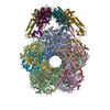
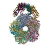
- EMDB-2679: Electron cryo-microscopy of the complex formed between the hexame... -
+ Open data

- Basic information
| Entry | Database: EMDB / ID: EMD-2679 | |||||||||
|---|---|---|---|---|---|---|---|---|---|---|
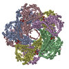
| Title | Electron cryo-microscopy of the complex formed between the hexameric AAA+ ATPase RavA and the decameric inducible decarboxylase LdcI | |||||||||
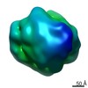
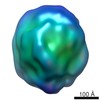
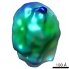
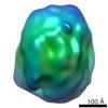
 Map data Map data | Cryo-EM reconstruction of the E. coli LdcI-RavA complex | |||||||||
 Sample Sample |
| |||||||||
 Keywords Keywords | Lysine decarboxylase / AAA+ ATPase / bacterial acid stress | |||||||||
| Function / homology |  Function and homology information Function and homology informationlysine decarboxylase / lysine decarboxylase activity / : / guanosine tetraphosphate binding / Hydrolases; Acting on acid anhydrides; In phosphorus-containing anhydrides / ATP hydrolysis activity / ATP binding / identical protein binding / plasma membrane / cytoplasm / cytosol Similarity search - Function | |||||||||
| Biological species |  | |||||||||
| Method | single particle reconstruction / cryo EM / Resolution: 11.0 Å | |||||||||
 Authors Authors | Malet H / Liu K / El Bakkouri M / Chan SWS / Effantin G / Bacia M / Houry WA / Gutsche I | |||||||||
 Citation Citation | Journal: Elife / Year: 2014 Title: Assembly principles of a unique cage formed by hexameric and decameric E. coli proteins. Authors: Hélène Malet / Kaiyin Liu / Majida El Bakkouri / Sze Wah Samuel Chan / Gregory Effantin / Maria Bacia / Walid A Houry / Irina Gutsche /   Abstract: A 3.3 MDa macromolecular cage between two Escherichia coli proteins with seemingly incompatible symmetries-the hexameric AAA+ ATPase RavA and the decameric inducible lysine decarboxylase LdcI-is ...A 3.3 MDa macromolecular cage between two Escherichia coli proteins with seemingly incompatible symmetries-the hexameric AAA+ ATPase RavA and the decameric inducible lysine decarboxylase LdcI-is reconstructed by cryo-electron microscopy to 11 Å resolution. Combined with a 7.5 Å resolution reconstruction of the minimal complex between LdcI and the LdcI-binding domain of RavA, and the previously solved crystal structures of the individual components, this work enables to build a reliable pseudoatomic model of this unusual architecture and to identify conformational rearrangements and specific elements essential for complex formation. The design of the cage created via lateral interactions between five RavA rings is unique for the diverse AAA+ ATPase superfamily. | |||||||||
| History |
|
- Structure visualization
| Movie |
Movie viewer |
|---|---|
| Structure viewer | EM map: SurfViewMolmilJmol/JSmol |
| Supplemental images |


- Downloads & links
-EMDB archive
| Map data | emd_2679.map.gz | 28.7 MB | EMDB map data format | |
|---|---|---|---|---|
| Header (meta data) | emd-2679-v30.xmlemd-2679.xml | 13.7 KB 13.7 KB | Display Display | EMDB header |
| Images |  EMDB-2679.png EMDB-2679.png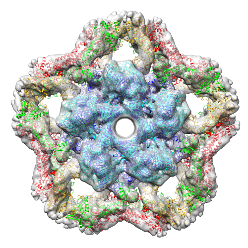 EMDB_2679.png EMDB_2679.png | 442.5 KB 442.5 KB | ||
| Archive directory |  http://ftp.pdbj.org/pub/emdb/structures/EMD-2679ftp://ftp.pdbj.org/pub/emdb/structures/EMD-2679 http://ftp.pdbj.org/pub/emdb/structures/EMD-2679ftp://ftp.pdbj.org/pub/emdb/structures/EMD-2679 | HTTPS FTP |
-Related structure data
| Related structure data | 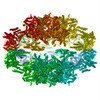 4upbMC 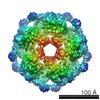 2681C  4upfC M: atomic model generated by this map C: citing same article ( |
|---|---|
| Similar structure data |
-Links
| EMDB pages | EMDB (EBI/PDBe) / EMDataResource |
|---|
-Map
| File | Download / File: emd_2679.map.gz / Format: CCP4 / Size: 29.8 MB / Type: IMAGE STORED AS FLOATING POINT NUMBER (4 BYTES) | ||||||||||||||||||||||||||||||||||||||||||||||||||||||||||||
|---|---|---|---|---|---|---|---|---|---|---|---|---|---|---|---|---|---|---|---|---|---|---|---|---|---|---|---|---|---|---|---|---|---|---|---|---|---|---|---|---|---|---|---|---|---|---|---|---|---|---|---|---|---|---|---|---|---|---|---|---|---|
| Annotation | Cryo-EM reconstruction of the E. coli LdcI-RavA complex | ||||||||||||||||||||||||||||||||||||||||||||||||||||||||||||
| Projections & slices | Image control
Images are generated by Spider. | ||||||||||||||||||||||||||||||||||||||||||||||||||||||||||||
| Voxel size | X=Y=Z: 2.37 Å | ||||||||||||||||||||||||||||||||||||||||||||||||||||||||||||
| Density |
| ||||||||||||||||||||||||||||||||||||||||||||||||||||||||||||
| Symmetry | Space group: 1 | ||||||||||||||||||||||||||||||||||||||||||||||||||||||||||||
| Details | EMDB XML:
CCP4 map header:
| ||||||||||||||||||||||||||||||||||||||||||||||||||||||||||||
 Z (Sec.)
 Y (Row.)
 X (Col.)


-Supplemental data
- Sample components
-Entire : Complex between the E.coli inducible lysine decarboxylase LdcI an...
| Entire | Name: Complex between the E.coli inducible lysine decarboxylase LdcI and the E. coli AAA+ ATPase RavA |
|---|---|
| Components |
|
-Supramolecule #1000: Complex between the E.coli inducible lysine decarboxylase LdcI an...
| Supramolecule | Name: Complex between the E.coli inducible lysine decarboxylase LdcI and the E. coli AAA+ ATPase RavA type: sample / ID: 1000 Oligomeric state: Two homodecamers of LdcI bind to five homohexamers of RavA Number unique components: 2 |
|---|---|
| Molecular weight | Experimental: 3.3 MDa / Theoretical: 3.3 MDa Method: Analytical ultracentrifugation Size exclusion chromatography |
-Macromolecule #1: Inducible lysine decarboxylase
| Macromolecule | Name: Inducible lysine decarboxylase / type: protein_or_peptide / ID: 1 / Name.synonym: LdcI / Number of copies: 20 / Oligomeric state: Two decamers / Recombinant expression: Yes |
|---|---|
| Source (natural) | Organism: |
| Molecular weight | Experimental: 81.2 KDa / Theoretical: 81.2 KDa |
| Recombinant expression | Organism: |
| Sequence | UniProtKB: Inducible lysine decarboxylase / GO: GO: 0006554 / InterPro: Ornithine/lysine/arginine decarboxylase |
-Macromolecule #2: AAA+ ATPase RavA
| Macromolecule | Name: AAA+ ATPase RavA / type: protein_or_peptide / ID: 2 / Name.synonym: RavA / Number of copies: 30 / Oligomeric state: Five homohexamers / Recombinant expression: Yes |
|---|---|
| Source (natural) | Organism: |
| Molecular weight | Experimental: 56.39 KDa / Theoretical: 56.39 KDa |
| Recombinant expression | Organism: |
| Sequence | UniProtKB: Regulatory ATPase RavA / GO: ATP hydrolysis activity / InterPro: ATPase RavA |
-Experimental details
-Structure determination
| Method | cryo EM |
|---|---|
 Processing Processing | single particle reconstruction |
| Aggregation state | particle |
-Sample preparation
| Concentration | 0.94 mg/mL |
|---|---|
| Buffer | pH: 6.5 Details: 25 mM MES pH 6.5, 200 mM NaCl, 3mM ADP, 0.8 mM PLP, 1mM DTT |
| Grid | Details: 400 mesh 2/1 1.2/1.3 quantifoil grids Glow discharged |
| Vitrification | Cryogen name: ETHANE / Chamber humidity: 100 % / Chamber temperature: 91 K / Instrument: FEI VITROBOT MARK III / Method: Blot 2 or 3 s before plunging |
- Electron microscopy
| Microscope | FEI POLARA 300 |
|---|---|
| Temperature | Min: 90 K / Max: 92 K / Average: 91 K |
| Alignment procedure | Legacy - Astigmatism: Objective lens astigmatism was corrected at 100,000 times magnification Legacy - Electron beam tilt params: 0 |
| Details | Weak beam illumination |
| Date | Apr 8, 2012 |
| Image recording | Category: FILM / Film or detector model: GATAN ULTRASCAN 4000 (4k x 4k) / Digitization - Scanner: ZEISS SCAI / Digitization - Sampling interval: 7 µm / Average electron dose: 20 e/Å2 / Bits/pixel: 8 |
| Tilt angle min | 0 |
| Tilt angle max | 0 |
| Electron beam | Acceleration voltage: 300 kV / Electron source:  FIELD EMISSION GUN FIELD EMISSION GUN |
| Electron optics | Calibrated magnification: 59000 / Illumination mode: FLOOD BEAM / Imaging mode: BRIGHT FIELD / Cs: 2 mm / Nominal defocus max: 3.3 µm / Nominal defocus min: 1.3 µm / Nominal magnification: 59000 |
| Sample stage | Specimen holder: Nitrogen cooled / Specimen holder model: GATAN HELIUM |
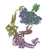
| Experimental equipment |  Model: Tecnai Polara / Image courtesy: FEI Company |
-Image processing
| Details | Initial model determined by cryo-ET and sub-tomogram averaging using IMOD and PEET. Projections of the initial model used for automated picking using the Fast Projection Matching algorithm. Model refined using SPIDER. |
|---|---|
| CTF correction | Details: Phase flipping |
| Final reconstruction | Applied symmetry - Point group: D5 (2x5 fold dihedral) / Algorithm: OTHER / Resolution.type: BY AUTHOR / Resolution: 11.0 Å / Resolution method: OTHER Software - Name: IMOD, PEET, CTFFIND3D, EMAN, (boxer), FPM, SPIDER Number images used: 21265 |
-Atomic model buiding 1
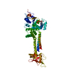
| Initial model | PDB ID: Chain - Chain ID: A |
|---|---|
| Software | Name: Flex-EM |
| Details | 3 rigid bodies, linkers between rigid bodies left flexible |
| Refinement | Space: REAL / Protocol: FLEXIBLE FIT / Target criteria: Cross-correlation, energy |
| Output model | PDB-4upb: |
-Atomic model buiding 2
| Initial model | PDB ID: Chain - Chain ID: X |
|---|---|
| Software | Name: Flex-EM |
| Details | 3 rigid bodies, linkers between rigid bodies left flexible |
| Refinement | Space: REAL / Protocol: FLEXIBLE FIT / Target criteria: Cross-correlation, energy |
| Output model | PDB-4upb: |