+ Open data
- Basic information
| Entry |  | |||||||||
|---|---|---|---|---|---|---|---|---|---|---|
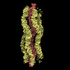
| Title | Cryo-EM of bundling pili from Pyrobaculum calidifontis | |||||||||
 Map data Map data | Cryo-EM and archaeal bundling filaments | |||||||||
 Sample Sample |
| |||||||||
 Keywords Keywords | helical symmetry / biofilm / bundling pili / pili / PROTEIN FIBRIL | |||||||||
| Function / homology | Uncharacterized protein Function and homology information Function and homology information | |||||||||
| Biological species |   Pyrobaculum calidifontis (archaea) Pyrobaculum calidifontis (archaea) | |||||||||
| Method | helical reconstruction / cryo EM / Resolution: 4.0 Å | |||||||||
 Authors Authors | Wang F / Cvirkaite-Krupovic V / Krupovic M / Egelman EH | |||||||||
| Funding support |  United States, 2 items United States, 2 items
| |||||||||
 Citation Citation |  Journal: Proc Natl Acad Sci U S A / Year: 2022 Journal: Proc Natl Acad Sci U S A / Year: 2022Title: Archaeal bundling pili of reveal similarities between archaeal and bacterial biofilms. Authors: Fengbin Wang / Virginija Cvirkaite-Krupovic / Mart Krupovic / Edward H Egelman /   Abstract: While biofilms formed by bacteria have received great attention due to their importance in pathogenesis, much less research has been focused on the biofilms formed by archaea. It has been known that ...While biofilms formed by bacteria have received great attention due to their importance in pathogenesis, much less research has been focused on the biofilms formed by archaea. It has been known that extracellular filaments in archaea, such as type IV pili, hami, and cannulae, play a part in the formation of archaeal biofilms. We have used cryo-electron microscopy to determine the atomic structure of a previously uncharacterized class of archaeal surface filaments from hyperthermophilic These filaments, which we call archaeal bundling pili (ABP), assemble into highly ordered bipolar bundles. The bipolar nature of these bundles most likely arises from the association of filaments from at least two different cells. The component protein, AbpA, shows homology, both at the sequence and structural level, to the bacterial protein TasA, a major component of the extracellular matrix in bacterial biofilms, contributing to biofilm stability. We show that AbpA forms very stable filaments in a manner similar to the donor-strand exchange of bacterial TasA fibers and chaperone-usher pathway pili where a β-strand from one subunit is incorporated into a β-sheet of the next subunit. Our results reveal likely mechanistic similarities and evolutionary connection between bacterial and archaeal biofilms, and suggest that there could be many other archaeal surface filaments that are as yet uncharacterized. | |||||||||
| History |
|
- Structure visualization
| Supplemental images |
|---|
- Downloads & links
-EMDB archive
| Map data |  emd_26474.map.gz emd_26474.map.gz | 20.1 MB |  EMDB map data format EMDB map data format | |
|---|---|---|---|---|
| Header (meta data) |  emd-26474-v30.xml emd-26474-v30.xml emd-26474.xml emd-26474.xml | 13.6 KB 13.6 KB | Display Display |  EMDB header EMDB header |
| Images |  emd_26474.png emd_26474.png | 91.1 KB | ||
| Filedesc metadata |  emd-26474.cif.gz emd-26474.cif.gz | 4.8 KB | ||
| Others |  emd_26474_half_map_1.map.gz emd_26474_half_map_1.map.gz emd_26474_half_map_2.map.gz emd_26474_half_map_2.map.gz | 318.5 MB 318.5 MB | ||
| Archive directory |  http://ftp.pdbj.org/pub/emdb/structures/EMD-26474 http://ftp.pdbj.org/pub/emdb/structures/EMD-26474 ftp://ftp.pdbj.org/pub/emdb/structures/EMD-26474 ftp://ftp.pdbj.org/pub/emdb/structures/EMD-26474 | HTTPS FTP |
-Validation report
| Summary document |  emd_26474_validation.pdf.gz emd_26474_validation.pdf.gz | 1 MB | Display |  EMDB validaton report EMDB validaton report |
|---|---|---|---|---|
| Full document |  emd_26474_full_validation.pdf.gz emd_26474_full_validation.pdf.gz | 1 MB | Display | |
| Data in XML |  emd_26474_validation.xml.gz emd_26474_validation.xml.gz | 17.3 KB | Display | |
| Data in CIF |  emd_26474_validation.cif.gz emd_26474_validation.cif.gz | 20.5 KB | Display | |
| Arichive directory |  https://ftp.pdbj.org/pub/emdb/validation_reports/EMD-26474 https://ftp.pdbj.org/pub/emdb/validation_reports/EMD-26474 ftp://ftp.pdbj.org/pub/emdb/validation_reports/EMD-26474 ftp://ftp.pdbj.org/pub/emdb/validation_reports/EMD-26474 | HTTPS FTP |
-Related structure data
| Related structure data |  7uegMC M: atomic model generated by this map C: citing same article ( |
|---|---|
| Similar structure data | Similarity search - Function & homology  F&H Search F&H Search |
- Links
Links
| EMDB pages |  EMDB (EBI/PDBe) / EMDB (EBI/PDBe) /  EMDataResource EMDataResource |
|---|
- Map
Map
| File |  Download / File: emd_26474.map.gz / Format: CCP4 / Size: 343 MB / Type: IMAGE STORED AS FLOATING POINT NUMBER (4 BYTES) Download / File: emd_26474.map.gz / Format: CCP4 / Size: 343 MB / Type: IMAGE STORED AS FLOATING POINT NUMBER (4 BYTES) | ||||||||||||||||||||||||||||||||||||
|---|---|---|---|---|---|---|---|---|---|---|---|---|---|---|---|---|---|---|---|---|---|---|---|---|---|---|---|---|---|---|---|---|---|---|---|---|---|
| Annotation | Cryo-EM and archaeal bundling filaments | ||||||||||||||||||||||||||||||||||||
| Projections & slices | Image control
Images are generated by Spider. | ||||||||||||||||||||||||||||||||||||
| Voxel size | X=Y=Z: 1.08 Å | ||||||||||||||||||||||||||||||||||||
| Density |
| ||||||||||||||||||||||||||||||||||||
| Symmetry | Space group: 1 | ||||||||||||||||||||||||||||||||||||
| Details | EMDB XML:
|
-Supplemental data
-Half map: half map A
| File | emd_26474_half_map_1.map | ||||||||||||
|---|---|---|---|---|---|---|---|---|---|---|---|---|---|
| Annotation | half map A | ||||||||||||
| Projections & Slices |
| ||||||||||||
| Density Histograms |
-Half map: half map b
| File | emd_26474_half_map_2.map | ||||||||||||
|---|---|---|---|---|---|---|---|---|---|---|---|---|---|
| Annotation | half map b | ||||||||||||
| Projections & Slices |
| ||||||||||||
| Density Histograms |
- Sample components
-Entire : extracellular bundling filaments
| Entire | Name: extracellular bundling filaments |
|---|---|
| Components |
|
-Supramolecule #1: extracellular bundling filaments
| Supramolecule | Name: extracellular bundling filaments / type: complex / ID: 1 / Parent: 0 / Macromolecule list: all |
|---|---|
| Source (natural) | Organism:   Pyrobaculum calidifontis (archaea) Pyrobaculum calidifontis (archaea) |
-Macromolecule #1: Pilin
| Macromolecule | Name: Pilin / type: protein_or_peptide / ID: 1 / Number of copies: 6 / Enantiomer: LEVO |
|---|---|
| Source (natural) | Organism:   Pyrobaculum calidifontis (archaea) Pyrobaculum calidifontis (archaea) |
| Molecular weight | Theoretical: 22.16798 KDa |
| Sequence | String: MARKKNYRPL IALAALAVAA LAMATLTFTN LTYWLINATL PPAMKYPGTD TTITRSDSSG YNRYVYVSYY YDPSTGYNVT RISIVGFTG DPTNYTNVLQ LCNKYYSGTL YAKLVAVGTV GTTNYESYIK DFRVYFVNPT TTPNYVQFQG TSVTQSATGS V SIGPGQCA ...String: MARKKNYRPL IALAALAVAA LAMATLTFTN LTYWLINATL PPAMKYPGTD TTITRSDSSG YNRYVYVSYY YDPSTGYNVT RISIVGFTG DPTNYTNVLQ LCNKYYSGTL YAKLVAVGTV GTTNYESYIK DFRVYFVNPT TTPNYVQFQG TSVTQSATGS V SIGPGQCA TVGAYVLVDP SLPTSARDGK TVIATYQVNV VFSTSP UniProtKB: Uncharacterized protein |
-Experimental details
-Structure determination
| Method | cryo EM |
|---|---|
 Processing Processing | helical reconstruction |
| Aggregation state | filament |
- Sample preparation
| Buffer | pH: 6 |
|---|---|
| Vitrification | Cryogen name: ETHANE |
- Electron microscopy
| Microscope | FEI TITAN KRIOS |
|---|---|
| Image recording | Film or detector model: GATAN K3 (6k x 4k) / Average electron dose: 50.0 e/Å2 |
| Electron beam | Acceleration voltage: 300 kV / Electron source:  FIELD EMISSION GUN FIELD EMISSION GUN |
| Electron optics | Illumination mode: FLOOD BEAM / Imaging mode: BRIGHT FIELD / Nominal defocus max: 2.5 µm / Nominal defocus min: 1.0 µm |
| Experimental equipment |  Model: Titan Krios / Image courtesy: FEI Company |
- Image processing
| Final reconstruction | Applied symmetry - Helical parameters - Δz: 32.785 Å Applied symmetry - Helical parameters - Δ&Phi: -77.404 ° Applied symmetry - Helical parameters - Axial symmetry: C1 (asymmetric) Resolution.type: BY AUTHOR / Resolution: 4.0 Å / Resolution method: FSC 0.143 CUT-OFF / Number images used: 204565 |
|---|---|
| Startup model | Type of model: NONE |
| Final angle assignment | Type: NOT APPLICABLE |
 Movie
Movie Controller


 Z (Sec.)
 Y (Row.)
 X (Col.)


