 ムービー
ムービー コントローラー

[日本語] English
 万見
万見- EMDB-25847: Cryo-EM structure of the 20S Alpha 3 Deletion proteasome core particle -
+ データを開く

- 基本情報
基本情報
| 登録情報 |  | |||||||||
|---|---|---|---|---|---|---|---|---|---|---|
| タイトル | Cryo-EM structure of the 20S Alpha 3 Deletion proteasome core particle | |||||||||
 マップデータ マップデータ | Final Map For 20S Alpha 3 Deletion | |||||||||
 試料 試料 |
| |||||||||
 キーワード キーワード | PI31 / Fub1 / core particle / HYDROLASE | |||||||||
| 機能・相同性 |  機能・相同性情報 機能・相同性情報proteasome core complex assembly / nuclear outer membrane-endoplasmic reticulum membrane network / Cross-presentation of soluble exogenous antigens (endosomes) / TNFR2 non-canonical NF-kB pathway / proteasomal ubiquitin-independent protein catabolic process / Ubiquitin Mediated Degradation of Phosphorylated Cdc25A / Regulation of PTEN stability and activity / KEAP1-NFE2L2 pathway / CDK-mediated phosphorylation and removal of Cdc6 / Neddylation ...proteasome core complex assembly / nuclear outer membrane-endoplasmic reticulum membrane network / Cross-presentation of soluble exogenous antigens (endosomes) / TNFR2 non-canonical NF-kB pathway / proteasomal ubiquitin-independent protein catabolic process / Ubiquitin Mediated Degradation of Phosphorylated Cdc25A / Regulation of PTEN stability and activity / KEAP1-NFE2L2 pathway / CDK-mediated phosphorylation and removal of Cdc6 / Neddylation / FBXL7 down-regulates AURKA during mitotic entry and in early mitosis / Orc1 removal from chromatin / MAPK6/MAPK4 signaling / proteasome storage granule / Antigen processing: Ubiquitination & Proteasome degradation / endopeptidase activator activity / proteasome assembly / proteasome endopeptidase complex / proteasome core complex, beta-subunit complex / proteasome core complex, alpha-subunit complex / threonine-type endopeptidase activity / Ub-specific processing proteases / Neutrophil degranulation / proteasome complex / proteasome-mediated ubiquitin-dependent protein catabolic process / endopeptidase activity / mRNA binding / endoplasmic reticulum membrane / mitochondrion / nucleus / cytosol 類似検索 - 分子機能 | |||||||||
| 生物種 |  | |||||||||
| 手法 | 単粒子再構成法 / クライオ電子顕微鏡法 / 解像度: 2.74 Å | |||||||||
 データ登録者 データ登録者 | Walsh Jr RM / Rawson S | |||||||||
| 資金援助 |  米国, 1件 米国, 1件
| |||||||||
 引用 引用 | ジャーナル: Nat Struct Mol Biol / 年: 2022 タイトル: Yeast PI31 inhibits the proteasome by a direct multisite mechanism. 著者: Shaun Rawson / Richard M Walsh / Benjamin Velez / Helena M Schnell / Fenglong Jiao / Marie Blickling / Jessie Ang / Meera K Bhanu / Lan Huang / John Hanna / 要旨: Proteasome inhibitors are widely used as therapeutics and research tools, and typically target one of the three active sites, each present twice in the proteasome complex. An endogeneous proteasome ...Proteasome inhibitors are widely used as therapeutics and research tools, and typically target one of the three active sites, each present twice in the proteasome complex. An endogeneous proteasome inhibitor, PI31, was identified 30 years ago, but its inhibitory mechanism has remained unclear. Here, we identify the mechanism of Saccharomyces cerevisiae PI31, also known as Fub1. Using cryo-electron microscopy (cryo-EM), we show that the conserved carboxy-terminal domain of Fub1 is present inside the proteasome's barrel-shaped core particle (CP), where it simultaneously interacts with all six active sites. Targeted mutations of Fub1 disrupt proteasome inhibition at one active site, while leaving the other sites unaffected. Fub1 itself evades degradation through distinct mechanisms at each active site. The gate that allows substrates to access the CP is constitutively closed, and Fub1 is enriched in mutant CPs with an abnormally open gate, suggesting that Fub1 may function to neutralize aberrant proteasomes, thereby ensuring the fidelity of proteasome-mediated protein degradation. | |||||||||
| 履歴 |
|
- 構造の表示
構造の表示
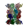
| 添付画像 |
|---|

- ダウンロードとリンク
-EMDBアーカイブ
| マップデータ | emd_25847.map.gz | 168.2 MB | EMDBマップデータ形式 | |
|---|---|---|---|---|
| ヘッダ (付随情報) | emd-25847-v30.xmlemd-25847.xml | 28 KB 28 KB | 表示 表示 | EMDBヘッダ |
| 画像 |  emd_25847.png emd_25847.png | 37.9 KB | ||
| Filedesc metadata | emd-25847.cif.gz | 8.2 KB | ||
| アーカイブディレクトリ |  http://ftp.pdbj.org/pub/emdb/structures/EMD-25847ftp://ftp.pdbj.org/pub/emdb/structures/EMD-25847 http://ftp.pdbj.org/pub/emdb/structures/EMD-25847ftp://ftp.pdbj.org/pub/emdb/structures/EMD-25847 | HTTPS FTP |
-検証レポート
| 文書・要旨 | emd_25847_validation.pdf.gz | 576.2 KB | 表示 | EMDB検証レポート |
|---|---|---|---|---|
| 文書・詳細版 | emd_25847_full_validation.pdf.gz | 575.7 KB | 表示 | |
| XML形式データ | emd_25847_validation.xml.gz | 7 KB | 表示 | |
| CIF形式データ | emd_25847_validation.cif.gz | 8 KB | 表示 | |
| アーカイブディレクトリ | https://ftp.pdbj.org/pub/emdb/validation_reports/EMD-25847ftp://ftp.pdbj.org/pub/emdb/validation_reports/EMD-25847 | HTTPS FTP |
-関連構造データ
-リンク
| EMDBのページ | EMDB (EBI/PDBe) / EMDataResource |
|---|---|
| 「今月の分子」の関連する項目 |


-マップ
| ファイル | ダウンロード / ファイル: emd_25847.map.gz / 形式: CCP4 / 大きさ: 178 MB / タイプ: IMAGE STORED AS FLOATING POINT NUMBER (4 BYTES) | ||||||||||||||||||||
|---|---|---|---|---|---|---|---|---|---|---|---|---|---|---|---|---|---|---|---|---|---|
| 注釈 | Final Map For 20S Alpha 3 Deletion | ||||||||||||||||||||
| ボクセルのサイズ | X=Y=Z: 1.06 Å | ||||||||||||||||||||
| 密度 |
| ||||||||||||||||||||
| 対称性 | 空間群: 1 | ||||||||||||||||||||
| 詳細 | EMDB XML:
|
-添付データ
- 試料の構成要素
+全体 : 20S proteasome from alpha3delta proteasome mutant
+超分子 #1: 20S proteasome from alpha3delta proteasome mutant
+分子 #1: Proteasome subunit beta type-6
+分子 #2: Proteasome subunit beta type-7
+分子 #3: Proteasome subunit alpha type-1
+分子 #4: Proteasome subunit alpha type-2
+分子 #5: Proteasome subunit alpha type-4
+分子 #6: Proteasome subunit alpha type-5
+分子 #7: Proteasome subunit alpha type-6
+分子 #8: Proteasome subunit alpha type-7
+分子 #9: Proteasome subunit beta type-1
+分子 #10: Proteasome subunit beta type-2
+分子 #11: Proteasome subunit beta type-3
+分子 #12: Proteasome subunit beta type-4
+分子 #13: Proteasome subunit beta type-5
-実験情報
-構造解析
| 手法 | クライオ電子顕微鏡法 |
|---|---|
 解析 解析 | 単粒子再構成法 |
| 試料の集合状態 | particle |
-試料調製
| 濃度 | 1.8 mg/mL | |||||||||||||||
|---|---|---|---|---|---|---|---|---|---|---|---|---|---|---|---|---|
| 緩衝液 | pH: 7.5 構成要素:
詳細: Fluorinated Fos-Choline was added to the sample immediately prior to deposition on a grid for plunge freezing. | |||||||||||||||
| グリッド | モデル: Quantifoil R1.2/1.3 / 材質: COPPER / メッシュ: 400 / 前処理 - タイプ: GLOW DISCHARGE / 前処理 - 時間: 30 sec. / 前処理 - 雰囲気: AIR / 前処理 - 気圧: 0.039 kPa / 詳細: 30 s glow discharge at 15 mA | |||||||||||||||
| 凍結 | 凍結剤: ETHANE / チャンバー内湿度: 100 % / チャンバー内温度: 295.15 K / 装置: FEI VITROBOT MARK IV |
- 電子顕微鏡法
| 顕微鏡 | FEI TITAN KRIOS |
|---|---|
| 特殊光学系 | エネルギーフィルター - 名称: GIF Bioquantum / エネルギーフィルター - スリット幅: 25 eV |
| 撮影 | フィルム・検出器のモデル: GATAN K3 BIOQUANTUM (6k x 4k) 撮影したグリッド数: 1 / 平均露光時間: 4.0 sec. / 平均電子線量: 53.85 e/Å2 |
| 電子線 | 加速電圧: 300 kV / 電子線源:  FIELD EMISSION GUN FIELD EMISSION GUN |
| 電子光学系 | C2レンズ絞り径: 50.0 µm / 倍率(補正後): 47169 / 照射モード: FLOOD BEAM / 撮影モード: BRIGHT FIELD / Cs: 2.7 mm / 最大 デフォーカス(公称値): 2.2 µm / 最小 デフォーカス(公称値): 0.8 µm / 倍率(公称値): 81000 |
| 試料ステージ | 試料ホルダーモデル: FEI TITAN KRIOS AUTOGRID HOLDER ホルダー冷却材: NITROGEN |
| 実験機器 |  モデル: Titan Krios / 画像提供: FEI Company |
+画像解析
-原子モデル構築 1
| 精密化 | 空間: REAL / プロトコル: AB INITIO MODEL |
|---|---|
| 得られたモデル | 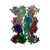 PDB-7tej: |
