[English] 日本語
Yorodumi
- EMDB-23868: Human N-type voltage-gated calcium channel Cav2.2 at 3.1 Angstrom... -

+
Open data


ID or keywords:

Loading...

-
Basic information

Entry
Database: EMDB / ID: EMD-23868
TitleHuman N-type voltage-gated calcium channel Cav2.2 at 3.1 Angstrom resolution
Map data
Sample
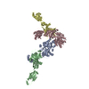
  • Complex: Cav2.2
    • Protein or peptide: Voltage-dependent N-type calcium channel subunit alpha-1B
    • Protein or peptide: Voltage-dependent L-type calcium channel subunit beta-3
    • Protein or peptide: Voltage-dependent calcium channel subunit alpha-2/delta-1
  • Ligand: 2-acetamido-2-deoxy-beta-D-glucopyranose
  • Ligand: CALCIUM ION
  • Ligand: CHOLESTEROL HEMISUCCINATE
  • Ligand: CHOLESTEROL
  • Ligand: 1,2-Distearoyl-sn-glycerophosphoethanolamine
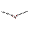
  • Ligand: [(2R)-1-octadecanoyloxy-3-[oxidanyl-[(1R,2R,3S,4R,5R,6S)-2,3,6-tris(oxidanyl)-4,5-diphosphonooxy-cyclohexyl]oxy-phospho ryl]oxy-propan-2-yl] (8Z)-icosa-5,8,11,14-tetraenoate
KeywordsCav2.2 / Channels / Calcium Ion-Selective / TRANSPORT PROTEIN
Function / homology
Function and homology information


regulation of membrane repolarization during action potential / Presynaptic depolarization and calcium channel opening / positive regulation of high voltage-gated calcium channel activity / calcium ion transmembrane transport via high voltage-gated calcium channel / membrane depolarization during bundle of His cell action potential / high voltage-gated calcium channel activity / L-type voltage-gated calcium channel complex / cardiac muscle cell action potential involved in contraction / NCAM1 interactions / regulation of ventricular cardiac muscle cell membrane repolarization ...regulation of membrane repolarization during action potential / Presynaptic depolarization and calcium channel opening / positive regulation of high voltage-gated calcium channel activity / calcium ion transmembrane transport via high voltage-gated calcium channel / membrane depolarization during bundle of His cell action potential / high voltage-gated calcium channel activity / L-type voltage-gated calcium channel complex / cardiac muscle cell action potential involved in contraction / NCAM1 interactions / regulation of ventricular cardiac muscle cell membrane repolarization / calcium ion transport into cytosol / regulation of calcium ion transmembrane transport via high voltage-gated calcium channel / voltage-gated calcium channel complex / neuromuscular junction development / calcium ion import across plasma membrane / neuronal dense core vesicle / regulation of heart rate by cardiac conduction / response to amyloid-beta / regulation of calcium ion transport / calcium channel regulator activity / voltage-gated calcium channel activity / sarcoplasmic reticulum / protein localization to plasma membrane / Regulation of insulin secretion / modulation of chemical synaptic transmission / Adrenaline,noradrenaline inhibits insulin secretion / cellular response to amyloid-beta / calcium ion transport / T cell receptor signaling pathway / amyloid-beta binding / chemical synaptic transmission / neuronal cell body / calcium ion binding / synapse / extracellular exosome / ATP binding / membrane / metal ion binding / plasma membrane / cytosol
Similarity search - Function
Voltage-dependent calcium channel, N-type, alpha-1 subunit / Voltage-dependent calcium channel, L-type, beta-3 subunit / Voltage-dependent L-type calcium channel subunit beta-3, SH3 domain / Voltage-dependent calcium channel, L-type, beta subunit / Voltage-dependent L-type calcium channel subunit beta-1-4, N-terminal A domain / Voltage gated calcium channel subunit beta domain 4Aa N terminal / VWA N-terminal / Voltage-dependent calcium channel, alpha-2/delta subunit, conserved region / : / VWA N-terminal ...Voltage-dependent calcium channel, N-type, alpha-1 subunit / Voltage-dependent calcium channel, L-type, beta-3 subunit / Voltage-dependent L-type calcium channel subunit beta-3, SH3 domain / Voltage-dependent calcium channel, L-type, beta subunit / Voltage-dependent L-type calcium channel subunit beta-1-4, N-terminal A domain / Voltage gated calcium channel subunit beta domain 4Aa N terminal / VWA N-terminal / Voltage-dependent calcium channel, alpha-2/delta subunit, conserved region / : / VWA N-terminal / Neuronal voltage-dependent calcium channel alpha 2acd / Voltage-dependent calcium channel, alpha-1 subunit, IQ domain / : / Voltage gated calcium channel IQ domain / Voltage gated calcium channel IQ domain / Voltage-dependent calcium channel, alpha-1 subunit / Voltage-dependent L-type calcium channel, IQ-associated domain / Voltage-dependent L-type calcium channel, IQ-associated / Guanylate kinase/L-type calcium channel beta subunit / Guanylate kinase / Guanylate kinase homologues. / von Willebrand factor type A domain / von Willebrand factor (vWF) type A domain / VWFA domain profile. / Voltage-dependent channel domain superfamily / von Willebrand factor, type A / von Willebrand factor A-like domain superfamily / EF-hand calcium-binding domain profile. / SH3-like domain superfamily / Src homology 3 (SH3) domain profile. / SH3 domain / EF-hand domain / Ion transport domain / Ion transport protein / P-loop containing nucleoside triphosphate hydrolase
Similarity search - Domain/homology
Voltage-dependent L-type calcium channel subunit beta-3 / Voltage-dependent calcium channel subunit alpha-2/delta-1 / Voltage-dependent N-type calcium channel subunit alpha-1B
Similarity search - Component
Biological speciesHomo sapiens (human)
Methodsingle particle reconstruction / cryo EM / Resolution: 3.1 Å
AuthorsYan N / Gao S
Funding support United States, 1 items
OrganizationGrant numberCountry
National Institutes of Health/National Institute of General Medical Sciences (NIH/NIGMS)5R01GM130762 United States
CitationJournal: Nature / Year: 2021
Title: Structure of human Ca2.2 channel blocked by the painkiller ziconotide.
Authors: Shuai Gao / Xia Yao / Nieng Yan /
Abstract: The neuronal-type (N-type) voltage-gated calcium (Ca) channels, which are designated Ca2.2, have an important role in the release of neurotransmitters. Ziconotide is a Ca2.2-specific peptide pore ...The neuronal-type (N-type) voltage-gated calcium (Ca) channels, which are designated Ca2.2, have an important role in the release of neurotransmitters. Ziconotide is a Ca2.2-specific peptide pore blocker that has been clinically used for treating intractable pain. Here we present cryo-electron microscopy structures of human Ca2.2 (comprising the core α1 and the ancillary α2δ-1 and β3 subunits) in the presence or absence of ziconotide. Ziconotide is thoroughly coordinated by helices P1 and P2, which support the selectivity filter, and the extracellular loops (ECLs) in repeats II, III and IV of α1. To accommodate ziconotide, the ECL of repeat III and α2δ-1 have to tilt upward concertedly. Three of the voltage-sensing domains (VSDs) are in a depolarized state, whereas the VSD of repeat II exhibits a down conformation that is stabilized by Ca2-unique intracellular segments and a phosphatidylinositol 4,5-bisphosphate molecule. Our studies reveal the molecular basis for Ca2.2-specific pore blocking by ziconotide and establish the framework for investigating electromechanical coupling in Ca channels.
History
DepositionApr 18, 2021-
Header (metadata) releaseJul 7, 2021-
Map releaseJul 7, 2021-
UpdateOct 30, 2024-
Current statusOct 30, 2024Processing site: RCSB / Status: Released

-
Structure visualization

Movie
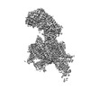
  • Surface view with section colored by density value
  • Surface level: 0.023
  • Imaged by UCSF Chimera
  • Download
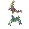
  • Surface view colored by height
  • Surface level: 0.023
  • Imaged by UCSF Chimera
  • Download
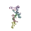
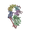
  • Surface view with fitted model
  • Atomic models: PDB-7miy
  • Surface level: 0.023
  • Imaged by UCSF Chimera
  • Download
Movie viewer
Structure viewerEM map:
SurfViewMolmilJmol/JSmol
Supplemental images

Downloads & links

-
Map

FileDownload / File: emd_23868.map.gz / Format: CCP4 / Size: 83.7 MB / Type: IMAGE STORED AS FLOATING POINT NUMBER (4 BYTES)
Projections & slices

Image control

Size
Brightness
Contrast
Others
AxesZ (Sec.)Y (Row.)X (Col.)
1.11 Å/pix.
x 280 pix.
= 311.92 Å
1.11 Å/pix.
x 280 pix.
= 311.92 Å
1.11 Å/pix.
x 280 pix.
= 311.92 Å

Surface

Projections

Slices (1/3)

Slices (1/2)

Slices (2/3)

Images are generated by Spider.

Voxel sizeX=Y=Z: 1.114 Å
Density
Contour LevelBy AUTHOR: 0.023 / Movie #1: 0.023
Minimum - Maximum-0.10476063 - 0.1651723
Average (Standard dev.)0.00001250235 (±0.0054037846)
SymmetrySpace group: 1
Details

EMDB XML:

Map geometry
Axis orderXYZ
Origin000
Dimensions280280280
Spacing280280280
CellA=B=C: 311.91998 Å
α=β=γ: 90.0 °

CCP4 map header:

modeImage stored as Reals
Å/pix. X/Y/Z1.1141.1141.114
M x/y/z280280280
origin x/y/z0.0000.0000.000
length x/y/z311.920311.920311.920
α/β/γ90.00090.00090.000
start NX/NY/NZ000
NX/NY/NZ256256256
MAP C/R/S123
start NC/NR/NS000
NC/NR/NS280280280
D min/max/mean-0.1050.1650.000

-
Supplemental data

-
Sample components

+
Entire : Cav2.2

EntireName: Cav2.2
Components
  • Complex: Cav2.2
    • Protein or peptide: Voltage-dependent N-type calcium channel subunit alpha-1B
    • Protein or peptide: Voltage-dependent L-type calcium channel subunit beta-3
    • Protein or peptide: Voltage-dependent calcium channel subunit alpha-2/delta-1
  • Ligand: 2-acetamido-2-deoxy-beta-D-glucopyranose
  • Ligand: CALCIUM ION
  • Ligand: CHOLESTEROL HEMISUCCINATE
  • Ligand: CHOLESTEROL
  • Ligand: 1,2-Distearoyl-sn-glycerophosphoethanolamine
  • Ligand: [(2R)-1-octadecanoyloxy-3-[oxidanyl-[(1R,2R,3S,4R,5R,6S)-2,3,6-tris(oxidanyl)-4,5-diphosphonooxy-cyclohexyl]oxy-phospho ryl]oxy-propan-2-yl] (8Z)-icosa-5,8,11,14-tetraenoate

+
Supramolecule #1: Cav2.2

SupramoleculeName: Cav2.2 / type: complex / ID: 1 / Parent: 0 / Macromolecule list: #1-#3
Source (natural)Organism: Homo sapiens (human)

+
Macromolecule #1: Voltage-dependent N-type calcium channel subunit alpha-1B

MacromoleculeName: Voltage-dependent N-type calcium channel subunit alpha-1B
type: protein_or_peptide / ID: 1 / Number of copies: 1 / Enantiomer: LEVO
Source (natural)Organism: Homo sapiens (human)
Molecular weightTheoretical: 262.831781 KDa
Recombinant expressionOrganism: Homo sapiens (human)
SequenceString: MVRFGDELGG RYGGPGGGER ARGGGAGGAG GPGPGGLQPG QRVLYKQSIA QRARTMALYN PIPVKQNCFT VNRSLFVFSE DNVVRKYAK RITEWPPFEY MILATIIANC IVLALEQHLP DGDKTPMSER LDDTEPYFIG IFCFEAGIKI IALGFVFHKG S YLRNGWNV ...String:
MVRFGDELGG RYGGPGGGER ARGGGAGGAG GPGPGGLQPG QRVLYKQSIA QRARTMALYN PIPVKQNCFT VNRSLFVFSE DNVVRKYAK RITEWPPFEY MILATIIANC IVLALEQHLP DGDKTPMSER LDDTEPYFIG IFCFEAGIKI IALGFVFHKG S YLRNGWNV MDFVVVLTGI LATAGTDFDL RTLRAVRVLR PLKLVSGIPS LQVVLKSIMK AMVPLLQIGL LLFFAILMFA II GLEFYMG KFHKACFPNS TDAEPVGDFP CGKEAPARLC EGDTECREYW PGPNFGITNF DNILFAILTV FQCITMEGWT DIL YNTNDA AGNTWNWLYF IPLIIIGSFF MLNLVLGVLS GEFAKERERV ENRRAFLKLR RQQQIERELN GYLEWIFKAE EVML AEEDR NAEEKSPLDV LKRAATKKSR NDLIHAEEGE DRFADLCAVG SPFARASLKS GKTESSSYFR RKEKMFRFFI RRMVK AQSF YWVVLCVVAL NTLCVAMVHY NQPRRLTTTL YFAEFVFLGL FLTEMSLKMY GLGPRSYFRS SFNCFDFGVI VGSVFE VVW AAIKPGSSFG ISVLRALRLL RIFKVTKYWS SLRNLVVSLL NSMKSIISLL FLLFLFIVVF ALLGMQLFGG QFNFQDE TP TTNFDTFPAA ILTVFQILTG EDWNAVMYHG IESQGGVSKG MFSSFYFIVL TLFGNYTLLN VFLAIAVDNL ANAQELTK D EEEMEEAANQ KLALQKAKEV AEVSPMSAAN ISIAARQQNS AKARSVWEQR ASQLRLQNLR ASCEALYSEM DPEERLRFA TTRHLRPDMK THLDRPLVVE LGRDGARGPV GGKARPEAAE APEGVDPPRR HHRHRDKDKT PAAGDQDRAE APKAESGEPG AREERPRPH RSHSKEAAGP PEARSERGRG PGPEGGRRHH RRGSPEEAAE REPRRHRAHR HQDPSKECAG AKGERRARHR G GPRAGPRE AESGEEPARR HRARHKAQPA HEAVEKETTE KEATEKEAEI VEADKEKELR NHQPREPHCD LETSGTVTVG PM HTLPSTC LQKVEEQPED ADNQRNVTRM GSQPPDPNTI VHIPVMLTGP LGEATVVPSG NVDLESQAEG KKEVEADDVM RSG PRPIVP YSSMFCLSPT NLLRRFCHYI VTMRYFEVVI LVVIALSSIA LAAEDPVRTD SPRNNALKYL DYIFTGVFTF EMVI KMIDL GLLLHPGAYF RDLWNILDFI VVSGALVAFA FSGSKGKDIN TIKSLRVLRV LRPLKTIKRL PKLKAVFDCV VNSLK NVLN ILIVYMLFMF IFAVIAVQLF KGKFFYCTDE SKELERDCRG QYLDYEKEEV EAQPRQWKKY DFHYDNVLWA LLTLFT VST GEGWPMVLKH SVDATYEEQG PSPGYRMELS IFYVVYFVVF PFFFVNIFVA LIIITFQEQG DKVMSECSLE KNERACI DF AISAKPLTRY MPQNRQSFQY KTWTFVVSPP FEYFIMAMIA LNTVVLMMKF YDAPYEYELM LKCLNIVFTS MFSMECVL K IIAFGVLNYF RDAWNVFDFV TVLGSITDIL VTEIAETNNF INLSFLRLFR AARLIKLLRQ GYTIRILLWT FVQSFKALP YVCLLIAMLF FIYAIIGMQV FGNIALDDDT SINRHNNFRT FLQALMLLFR SATGEAWHEI MLSCLSNQAC DEQANATECG SDFAYFYFV SFIFLCSFLM LNLFVAVIMD NFEYLTRDSS ILGPHHLDEF IRVWAEYDPA ACGRISYNDM FEMLKHMSPP L GLGKKCPA RVAYKRLVRM NMPISNEDMT VHFTSTLMAL IRTALEIKLA PAGTKQHQCD AELRKEISVV WANLPQKTLD LL VPPHKPD EMTVGKVYAA LMIFDFYKQN KTTRDQMQQA PGGLSQMGPV SLFHPLKATL EQTQPAVLRG ARVFLRQKSS TSL SNGGAI QNQESGIKES VSWGTQRTQD APHEARPPLE RGHSTEIPVG RSGALAVDVQ MQSITRRGPD GEPQPGLESQ GRAA SMPRL AAETQPVTDA SPMKRSISTL AQRPRGTHLC STTPDRPPPS QASSHHHHHR CHRRRDRKQR SLEKGPSLSA DMDGA PSSA VGPGLPPGEG PTGCRRERER RQERGRSQER RQPSSSSSEK QRFYSCDRFG GREPPKPKPS LSSHPTSPTA GQEPGP HPQ GSGSVNGSPL LSTSGASTPG RGGRRQLPQT PLTPRPSITY KTANSSPIHF AGAQTSLPAF SPGRLSRGLS EHNALLQ RD PLSQPLAPGS RIGSDPYLGQ RLDSEASVHA LPEDTLTFEE AVATNSGRSS RTSYVSSLTS QSHPLRRVPN GYHCTLGL S SGGRARHSYH HPDQDHWC

UniProtKB: Voltage-dependent N-type calcium channel subunit alpha-1B

+
Macromolecule #2: Voltage-dependent L-type calcium channel subunit beta-3

MacromoleculeName: Voltage-dependent L-type calcium channel subunit beta-3
type: protein_or_peptide / ID: 2 / Number of copies: 1 / Enantiomer: LEVO
Source (natural)Organism: Homo sapiens (human)
Molecular weightTheoretical: 54.607852 KDa
Recombinant expressionOrganism: Homo sapiens (human)
SequenceString: MYDDSYVPGF EDSEAGSADS YTSRPSLDSD VSLEEDRESA RREVESQAQQ QLERAKHKPV AFAVRTNVSY CGVLDEECPV QGSGVNFEA KDFLHIKEKY SNDWWIGRLV KEGGDIAFIP SPQRLESIRL KQEQKARRSG NPSSLSDIGN RRSPPPSLAK Q KQKQAEHV ...String:
MYDDSYVPGF EDSEAGSADS YTSRPSLDSD VSLEEDRESA RREVESQAQQ QLERAKHKPV AFAVRTNVSY CGVLDEECPV QGSGVNFEA KDFLHIKEKY SNDWWIGRLV KEGGDIAFIP SPQRLESIRL KQEQKARRSG NPSSLSDIGN RRSPPPSLAK Q KQKQAEHV PPYDVVPSMR PVVLVGPSLK GYEVTDMMQK ALFDFLKHRF DGRISITRVT ADLSLAKRSV LNNPGKRTII ER SSARSSI AEVQSEIERI FELAKSLQLV VLDADTINHP AQLAKTSLAP IIVFVKVSSP KVLQRLIRSR GKSQMKHLTV QMM AYDKLV QCPPESFDVI LDENQLEDAC EHLAEYLEVY WRATHHPAPG PGLLGPPSAI PGLQNQQLLG ERGEEHSPLE RDSL MPSDE ASESSRQAWT GSSQRSSRHL EEDYADAYQD LYQPHRQHTS GLPSANGHDP QDRLLAQDSE HNHSDRNWQR NRPWP KDSY

UniProtKB: Voltage-dependent L-type calcium channel subunit beta-3

+
Macromolecule #3: Voltage-dependent calcium channel subunit alpha-2/delta-1

MacromoleculeName: Voltage-dependent calcium channel subunit alpha-2/delta-1
type: protein_or_peptide / ID: 3 / Number of copies: 1 / Enantiomer: LEVO
Source (natural)Organism: Homo sapiens (human)
Molecular weightTheoretical: 124.692469 KDa
Recombinant expressionOrganism: Homo sapiens (human)
SequenceString: MAAGCLLALT LTLFQSLLIG PSSEEPFPSA VTIKSWVDKM QEDLVTLAKT ASGVNQLVDI YEKYQDLYTV EPNNARQLVE IAARDIEKL LSNRSKALVR LALEAEKVQA AHQWREDFAS NEVVYYNAKD DLDPEKNDSE PGSQRIKPVF IEDANFGRQI S YQHAAVHI ...String:
MAAGCLLALT LTLFQSLLIG PSSEEPFPSA VTIKSWVDKM QEDLVTLAKT ASGVNQLVDI YEKYQDLYTV EPNNARQLVE IAARDIEKL LSNRSKALVR LALEAEKVQA AHQWREDFAS NEVVYYNAKD DLDPEKNDSE PGSQRIKPVF IEDANFGRQI S YQHAAVHI PTDIYEGSTI VLNELNWTSA LDEVFKKNRE EDPSLLWQVF GSATGLARYY PASPWVDNSR TPNKIDLYDV RR RPWYIQG AASPKDMLIL VDVSGSVSGL TLKLIRTSVS EMLETLSDDD FVNVASFNSN AQDVSCFQHL VQANVRNKKV LKD AVNNIT AKGITDYKKG FSFAFEQLLN YNVSRANCNK IIMLFTDGGE ERAQEIFNKY NKDKKVRVFT FSVGQHNYDR GPIQ WMACE NKGYYYEIPS IGAIRINTQE YLDVLGRPMV LAGDKAKQVQ WTNVYLDALE LGLVITGTLP VFNITGQFEN KTNLK NQLI LGVMGVDVSL EDIKRLTPRF TLCPNGYYFA IDPNGYVLLH PNLQPKPIGV GIPTINLRKR RPNIQNPKSQ EPVTLD FLD AELENDIKVE IRNKMIDGES GEKTFRTLVK SQDERYIDKG NRTYTWTPVN GTDYSLALVL PTYSFYYIKA KLEETIT QA RYSETLKPDN FEESGYTFIA PRDYCNDLKI SDNNTEFLLN FNEFIDRKTP NNPSCNADLI NRVLLDAGFT NELVQNYW S KQKNIKGVKA RFVVTDGGIT RVYPKEAGEN WQENPETYED SFYKRSLDND NYVFTAPYFN KSGPGAYESG IMVSKAVEI YIQGKLLKPA VVGIKIDVNS WIENFTKTSI RDPCAGPVCD CKRNSDVMDC VILDDGGFLL MANHDDYTNQ IGRFFGEIDP SLMRHLVNI SVYAFNKSYD YQSVCEPGAA PKQGAGHRSA YVPSVADILQ IGWWATAAAW SILQQFLLSL TFPRLLEAVE M EDDDFTAS LSKQSCITEQ TQYFFDNDSK SFSGVLDCGN CSRIFHGEKL MNTNLIFIMV ESKGTCPCDT RLLIQAEQTS DG PNPCDMV KQPRYRKGPD VCFDNNVLED YTDCGGVSGL NPSLWYIIGI QFLLLWLVSG STHRLL

UniProtKB: Voltage-dependent calcium channel subunit alpha-2/delta-1

+
Macromolecule #7: 2-acetamido-2-deoxy-beta-D-glucopyranose

MacromoleculeName: 2-acetamido-2-deoxy-beta-D-glucopyranose / type: ligand / ID: 7 / Number of copies: 3 / Formula: NAG
Molecular weightTheoretical: 221.208 Da
Chemical component information

ChemComp-NAG:
2-acetamido-2-deoxy-beta-D-glucopyranose

+
Macromolecule #8: CALCIUM ION

MacromoleculeName: CALCIUM ION / type: ligand / ID: 8 / Number of copies: 2 / Formula: CA
Molecular weightTheoretical: 40.078 Da

+
Macromolecule #9: CHOLESTEROL HEMISUCCINATE

MacromoleculeName: CHOLESTEROL HEMISUCCINATE / type: ligand / ID: 9 / Number of copies: 3 / Formula: Y01
Molecular weightTheoretical: 486.726 Da
Chemical component information

ChemComp-Y01:
CHOLESTEROL HEMISUCCINATE

+
Macromolecule #10: CHOLESTEROL

MacromoleculeName: CHOLESTEROL / type: ligand / ID: 10 / Number of copies: 4 / Formula: CLR
Molecular weightTheoretical: 386.654 Da
Chemical component information

ChemComp-CLR:
CHOLESTEROL

+
Macromolecule #11: 1,2-Distearoyl-sn-glycerophosphoethanolamine

MacromoleculeName: 1,2-Distearoyl-sn-glycerophosphoethanolamine / type: ligand / ID: 11 / Number of copies: 2 / Formula: 3PE
Molecular weightTheoretical: 748.065 Da
Chemical component information

ChemComp-3PE:
1,2-Distearoyl-sn-glycerophosphoethanolamine / phospholipid*YM

+
Macromolecule #12: [(2R)-1-octadecanoyloxy-3-[oxidanyl-[(1R,2R,3S,4R,5R,6S)-2,3,6-tr...

MacromoleculeName: [(2R)-1-octadecanoyloxy-3-[oxidanyl-[(1R,2R,3S,4R,5R,6S)-2,3,6-tris(oxidanyl)-4,5-diphosphonooxy-cyclohexyl]oxy-phospho ryl]oxy-propan-2-yl] (8Z)-icosa-5,8,11,14-tetraenoate
type: ligand / ID: 12 / Number of copies: 1 / Formula: PT5
Molecular weightTheoretical: 1.047088 KDa

-
Experimental details

-
Structure determination

Methodcryo EM
Processingsingle particle reconstruction
Aggregation stateparticle

-
Sample preparation

Concentration20 mg/mL
BufferpH: 7.4
VitrificationCryogen name: ETHANE / Chamber humidity: 100 % / Chamber temperature: 281 K / Instrument: FEI VITROBOT MARK IV / Details: blot for 6 seconds before plunging.
DetailsCav2.2

-
Electron microscopy

MicroscopeFEI TITAN KRIOS
Image recordingFilm or detector model: GATAN K2 SUMMIT (4k x 4k) / Average electron dose: 50.0 e/Å2
Electron beamAcceleration voltage: 300 kV / Electron source: FIELD EMISSION GUN
Electron opticsIllumination mode: FLOOD BEAM / Imaging mode: BRIGHT FIELD
Experimental equipment
Model: Titan Krios / Image courtesy: FEI Company

-
Image processing

Startup modelType of model: OTHER
Final reconstructionResolution.type: BY AUTHOR / Resolution: 3.1 Å / Resolution method: FSC 0.143 CUT-OFF / Number images used: 56615
Initial angle assignmentType: MAXIMUM LIKELIHOOD
Final angle assignmentType: MAXIMUM LIKELIHOOD

+
About Yorodumi

-
News

-
Feb 9, 2022. New format data for meta-information of EMDB entries

New format data for meta-information of EMDB entries

  • Version 3 of the EMDB header file is now the official format.
  • The previous official version 1.9 will be removed from the archive.

Related info.:EMDB header

External links:wwPDB to switch to version 3 of the EMDB data model

-
Aug 12, 2020. Covid-19 info

Covid-19 info

URL: https://pdbj.org/emnavi/covid19.php

New page: Covid-19 featured information page in EM Navigator.

Related info.:Covid-19 info / Mar 5, 2020. Novel coronavirus structure data

+
Mar 5, 2020. Novel coronavirus structure data

Novel coronavirus structure data

Related info.:Yorodumi Speices / Aug 12, 2020. Covid-19 info

External links:COVID-19 featured content - PDBj / Molecule of the Month (242):Coronavirus Proteases

+
Jan 31, 2019. EMDB accession codes are about to change! (news from PDBe EMDB page)

EMDB accession codes are about to change! (news from PDBe EMDB page)

  • The allocation of 4 digits for EMDB accession codes will soon come to an end. Whilst these codes will remain in use, new EMDB accession codes will include an additional digit and will expand incrementally as the available range of codes is exhausted. The current 4-digit format prefixed with “EMD-” (i.e. EMD-XXXX) will advance to a 5-digit format (i.e. EMD-XXXXX), and so on. It is currently estimated that the 4-digit codes will be depleted around Spring 2019, at which point the 5-digit format will come into force.
  • The EM Navigator/Yorodumi systems omit the EMD- prefix.

Related info.:Q: What is EMD? / ID/Accession-code notation in Yorodumi/EM Navigator

External links:EMDB Accession Codes are Changing Soon! / Contact to PDBj

+
Jul 12, 2017. Major update of PDB

Major update of PDB

  • wwPDB released updated PDB data conforming to the new PDBx/mmCIF dictionary.
  • This is a major update changing the version number from 4 to 5, and with Remediation, in which all the entries are updated.
  • In this update, many items about electron microscopy experimental information are reorganized (e.g. em_software).
  • Now, EM Navigator and Yorodumi are based on the updated data.

External links:wwPDB Remediation / Enriched Model Files Conforming to OneDep Data Standards Now Available in the PDB FTP Archive

-
Yorodumi

Thousand views of thousand structures

  • Yorodumi is a browser for structure data from EMDB, PDB, SASBDB, etc.
  • This page is also the successor to EM Navigator detail page, and also detail information page/front-end page for Omokage search.
  • The word "yorodu" (or yorozu) is an old Japanese word meaning "ten thousand". "mi" (miru) is to see.

Related info.:EMDB / PDB / SASBDB / Comparison of 3 databanks / Yorodumi Search / Aug 31, 2016. New EM Navigator & Yorodumi / Yorodumi Papers / Jmol/JSmol / Function and homology information / Changes in new EM Navigator and Yorodumi

Read more