 Movie
Movie Controller

[English] 日本語
 Yorodumi
- EMDB-21380: Structure of an acid-sensing ion channel solubilized by styrene m... -
+ Open data

- Basic information
| Entry | Database: EMDB / ID: EMD-21380 | ||||||||||||
|---|---|---|---|---|---|---|---|---|---|---|---|---|---|
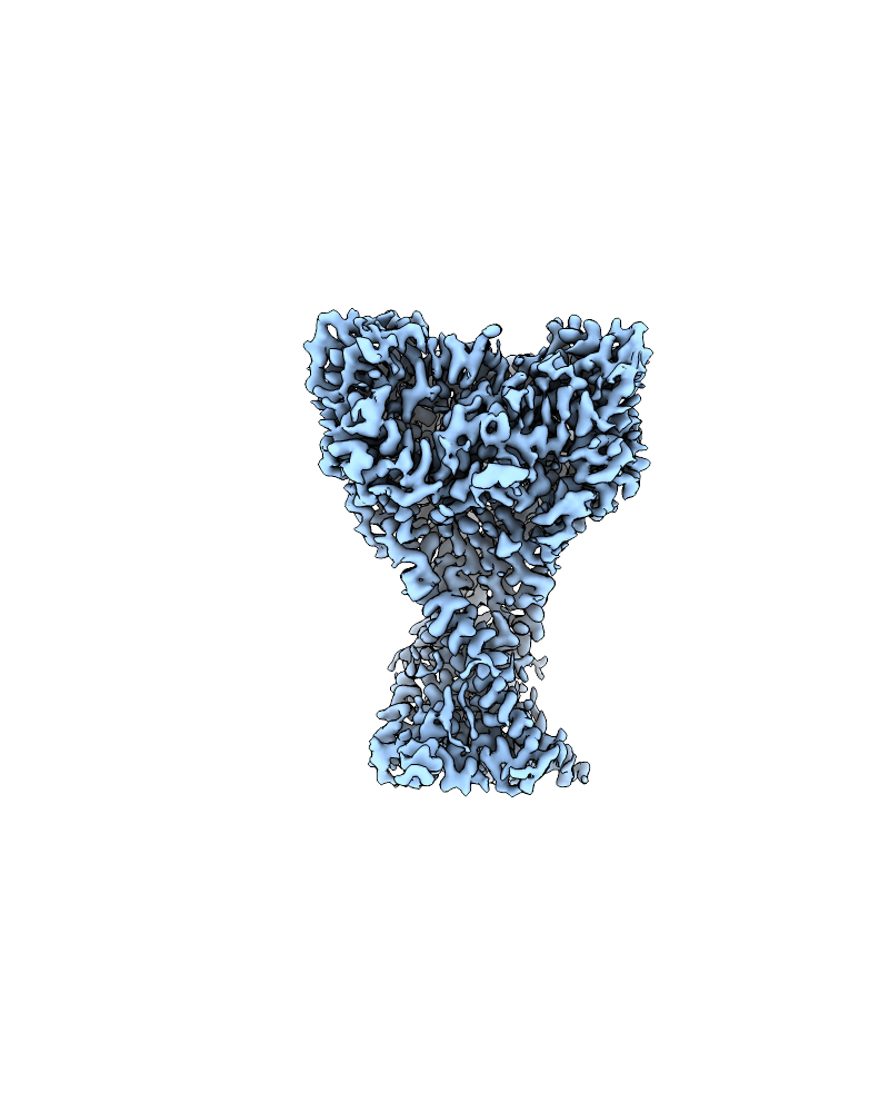
| Title | Structure of an acid-sensing ion channel solubilized by styrene maleic acid and in a desensitized state at low pH | ||||||||||||
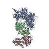
 Map data Map data | Map of SMA-cASIC1a in a desensitized state at low pH. | ||||||||||||
 Sample Sample |
| ||||||||||||
 Keywords Keywords | ion channel / ligand-gated ion channel / acid-sensing ion channel / ASIC / ASIC1a / proton-gated ion channel / membrane protein / TRANSPORT PROTEIN | ||||||||||||
| Function / homology |  Function and homology information Function and homology informationStimuli-sensing channels / pH-gated monoatomic ion channel activity / cellular response to pH / neurotransmitter secretion / ligand-gated sodium channel activity / sodium ion transmembrane transport / presynapse / postsynaptic membrane / dendrite / glutamatergic synapse ...Stimuli-sensing channels / pH-gated monoatomic ion channel activity / cellular response to pH / neurotransmitter secretion / ligand-gated sodium channel activity / sodium ion transmembrane transport / presynapse / postsynaptic membrane / dendrite / glutamatergic synapse / identical protein binding / plasma membrane Similarity search - Function | ||||||||||||
| Biological species |  | ||||||||||||
| Method | single particle reconstruction / cryo EM / Resolution: 2.82 Å | ||||||||||||
 Authors Authors | Yoder N / Gouaux E | ||||||||||||
| Funding support |  United States, 3 items United States, 3 items
| ||||||||||||
 Citation Citation | Journal: Elife / Year: 2020 Title: The His-Gly motif of acid-sensing ion channels resides in a reentrant 'loop' implicated in gating and ion selectivity. Authors: Nate Yoder / Eric Gouaux / Abstract: Acid-sensing ion channels (ASICs) are proton-gated members of the epithelial sodium channel/degenerin (ENaC/DEG) superfamily of ion channels and are expressed throughout the central and peripheral ...Acid-sensing ion channels (ASICs) are proton-gated members of the epithelial sodium channel/degenerin (ENaC/DEG) superfamily of ion channels and are expressed throughout the central and peripheral nervous systems. The homotrimeric splice variant ASIC1a has been implicated in nociception, fear memory, mood disorders and ischemia. Here, we extract full-length chicken ASIC1 (cASIC1) from cell membranes using styrene maleic acid (SMA) copolymer, elucidating structures of ASIC1 channels in both high pH resting and low pH desensitized conformations by single-particle cryo-electron microscopy (cryo-EM). The structures of resting and desensitized channels reveal a reentrant loop at the amino terminus of ASIC1 that includes the highly conserved 'His-Gly' (HG) motif. The reentrant loop lines the lower ion permeation pathway and buttresses the 'Gly-Ala-Ser' (GAS) constriction, thus providing a structural explanation for the role of the His-Gly dipeptide in the structure and function of ASICs. #1: Journal: Biorxiv / Year: 2020Title: Conserved His-Gly motif of acid-sensing ion channels resides in a reentrant loop implicated in gating and ion selectivity Authors: Yoder N / Gouaux E | ||||||||||||
| History |
|
- Structure visualization
| Movie |
Movie viewer |
|---|---|
| Structure viewer | EM map: SurfViewMolmilJmol/JSmol |
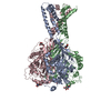
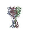
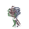
| Supplemental images |
- Downloads & links
-EMDB archive
| Map data | emd_21380.map.gz | 168 MB | EMDB map data format | |
|---|---|---|---|---|
| Header (meta data) | emd-21380-v30.xmlemd-21380.xml | 13.2 KB 13.2 KB | Display Display | EMDB header |
| Images |  emd_21380.png emd_21380.png | 231.3 KB | ||
| Filedesc metadata | emd-21380.cif.gz | 5.9 KB | ||
| Archive directory |  http://ftp.pdbj.org/pub/emdb/structures/EMD-21380ftp://ftp.pdbj.org/pub/emdb/structures/EMD-21380 http://ftp.pdbj.org/pub/emdb/structures/EMD-21380ftp://ftp.pdbj.org/pub/emdb/structures/EMD-21380 | HTTPS FTP |
-Related structure data
| Related structure data |  6vtkMC  6vtlC M: atomic model generated by this map C: citing same article ( |
|---|---|
| Similar structure data |


-Links
| EMDB pages | EMDB (EBI/PDBe) / EMDataResource |
|---|
-Map
| File | Download / File: emd_21380.map.gz / Format: CCP4 / Size: 178 MB / Type: IMAGE STORED AS FLOATING POINT NUMBER (4 BYTES) | ||||||||||||||||||||||||||||||||||||||||||||||||||||||||||||||||||||
|---|---|---|---|---|---|---|---|---|---|---|---|---|---|---|---|---|---|---|---|---|---|---|---|---|---|---|---|---|---|---|---|---|---|---|---|---|---|---|---|---|---|---|---|---|---|---|---|---|---|---|---|---|---|---|---|---|---|---|---|---|---|---|---|---|---|---|---|---|---|
| Annotation | Map of SMA-cASIC1a in a desensitized state at low pH. | ||||||||||||||||||||||||||||||||||||||||||||||||||||||||||||||||||||
| Projections & slices | Image control
Images are generated by Spider. | ||||||||||||||||||||||||||||||||||||||||||||||||||||||||||||||||||||
| Voxel size | X=Y=Z: 1.096 Å | ||||||||||||||||||||||||||||||||||||||||||||||||||||||||||||||||||||
| Density |
| ||||||||||||||||||||||||||||||||||||||||||||||||||||||||||||||||||||
| Symmetry | Space group: 1 | ||||||||||||||||||||||||||||||||||||||||||||||||||||||||||||||||||||
| Details | EMDB XML:
CCP4 map header:
| ||||||||||||||||||||||||||||||||||||||||||||||||||||||||||||||||||||
 Z (Sec.)
 Y (Row.)
 X (Col.)


-Supplemental data
- Sample components
-Entire : Acid-sensing ion channel
| Entire | Name: Acid-sensing ion channel |
|---|---|
| Components |
|
-Supramolecule #1: Acid-sensing ion channel
| Supramolecule | Name: Acid-sensing ion channel / type: complex / ID: 1 / Parent: 0 / Macromolecule list: #1 |
|---|---|
| Source (natural) | Organism: |
| Molecular weight | Theoretical: 180 KDa |
-Macromolecule #1: Acid-sensing ion channel 1
| Macromolecule | Name: Acid-sensing ion channel 1 / type: protein_or_peptide / ID: 1 / Number of copies: 3 / Enantiomer: LEVO |
|---|---|
| Source (natural) | Organism: |
| Molecular weight | Theoretical: 60.080324 KDa |
| Recombinant expression | Organism:  Homo sapiens (human) Homo sapiens (human) |
| Sequence | String: MMDLKVDEEE VDSGQPVSIQ AFASSSTLHG ISHIFSYERL SLKRVVWALC FMGSLALLAL VCTNRIQYYF LYPHVTKLDE VAATRLTFP AVTFCNLNEF RFSRVTKNDL YHAGELLALL NNRYEIPDTQ TADEKQLEIL QDKANFRNFK PKPFNMLEFY D RAGHDIRE ...String: MMDLKVDEEE VDSGQPVSIQ AFASSSTLHG ISHIFSYERL SLKRVVWALC FMGSLALLAL VCTNRIQYYF LYPHVTKLDE VAATRLTFP AVTFCNLNEF RFSRVTKNDL YHAGELLALL NNRYEIPDTQ TADEKQLEIL QDKANFRNFK PKPFNMLEFY D RAGHDIRE MLLSCFFRGE QCSPEDFKVV FTRYGKCYTF NAGQDGKPRL ITMKGGTGNG LEIMLDIQQD EYLPVWGETD ET SFEAGIK VQIHSQDEPP LIDQLGFGVA PGFQTFVSCQ EQRLIYLPPP WGDCKATTGD SEFYDTYSIT ACRIDCETRY LVE NCNCRM VHMPGDAPYC TPEQYKECAD PALDFLVEKD NEYCVCEMPC NVTRYGKELS MVKIPSKASA KYLAKKYNKS EQYI GENIL VLDIFFEALN YETIEQKKAY EVAGLLGDIG GQMGLFIGAS ILTVLELFDY AYEVIKHRLC RRGKCRKNHK RNNTD KGVA LSMDDVKRHN PCESLRGHPA GMTYAANILP HHPARGTFED FTC UniProtKB: Acid-sensing ion channel 1 |
-Macromolecule #2: 2-acetamido-2-deoxy-beta-D-glucopyranose
| Macromolecule | Name: 2-acetamido-2-deoxy-beta-D-glucopyranose / type: ligand / ID: 2 / Number of copies: 6 / Formula: NAG |
|---|---|
| Molecular weight | Theoretical: 221.208 Da |
| Chemical component information |  ChemComp-NAG: |
-Experimental details
-Structure determination
| Method | cryo EM |
|---|---|
 Processing Processing | single particle reconstruction |
| Aggregation state | particle |
-Sample preparation
| Concentration | 1.00 mg/mL |
|---|---|
| Buffer | pH: 7 |
| Grid | Model: Quantifoil R1.2/1.3 / Material: GOLD / Mesh: 200 / Pretreatment - Type: GLOW DISCHARGE / Pretreatment - Time: 30 sec. / Pretreatment - Atmosphere: AIR |
| Vitrification | Cryogen name: ETHANE-PROPANE / Chamber humidity: 70 % / Chamber temperature: 277.15 K / Instrument: HOMEMADE PLUNGER Details: Specimen was frozen on a custom-made manual plunge apparatus in a cold room environment maintained at high humidity.. |
- Electron microscopy
| Microscope | FEI TITAN KRIOS |
|---|---|
| Image recording | Film or detector model: GATAN K2 SUMMIT (4k x 4k) / Detector mode: SUPER-RESOLUTION / Average electron dose: 50.0 e/Å2 |
| Electron beam | Acceleration voltage: 300 kV / Electron source:  FIELD EMISSION GUN FIELD EMISSION GUN |
| Electron optics | Illumination mode: FLOOD BEAM / Imaging mode: BRIGHT FIELD |
| Experimental equipment |  Model: Titan Krios / Image courtesy: FEI Company |
-Image processing
| Startup model | Type of model: NONE |
|---|---|
| Final reconstruction | Applied symmetry - Point group: C3 (3 fold cyclic) / Resolution.type: BY AUTHOR / Resolution: 2.82 Å / Resolution method: FSC 0.143 CUT-OFF / Software - Name: cryoSPARC (ver. 2) / Number images used: 121148 |
| Initial angle assignment | Type: RANDOM ASSIGNMENT / Software - Name: cryoSPARC (ver. 2) |
| Final angle assignment | Type: MAXIMUM LIKELIHOOD / Software - Name: cryoSPARC (ver. 2) |

