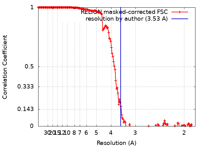
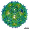
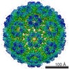
Journal: PLoS Comput Biol / Year: 2020 Title: Expression of quasi-equivalence and capsid dimorphism in the Hepadnaviridae. Authors: Weimin Wu / Norman R Watts / Naiqian Cheng / Rick Huang / Alasdair C Steven / Paul T Wingfield / Abstract: Hepatitis B virus (HBV) is a leading cause of liver disease. The capsid is an essential component of the virion and it is therefore of interest how it assembles and disassembles. The capsid protein ...Hepatitis B virus (HBV) is a leading cause of liver disease. The capsid is an essential component of the virion and it is therefore of interest how it assembles and disassembles. The capsid protein is unusual both for its rare fold and that it polymerizes according to two different icosahedral symmetries, causing the polypeptide chain to exist in seven quasi-equivalent environments: A, B, and C in AB and CC dimers in T = 3 capsids, and A, B, C, and D in AB and CD dimers in T = 4 capsids. We have compared the two capsids by cryo-EM at 3.5 Å resolution. To ensure a valid comparison, the two capsids were prepared and imaged under identical conditions. We find that the chains have different conformations and potential energies, with the T = 3 C chain having the lowest. Three of the four quasi-equivalent dimers are asymmetric with respect to conformation and potential energy; however, the T = 3 CC dimer is symmetrical and has the lowest potential energy although its intra-dimer interface has the least free energy of formation. Of all the inter-dimer interfaces, the CB interface has the least area and free energy, in both capsids. From the calculated energies of higher-order groupings of dimers discernible in the lattices we predict early assembly intermediates, and indeed we observe such structures by negative stain EM of in vitro assembly reactions. By sequence analysis and computational alanine scanning we identify key residues and motifs involved in capsid assembly. Our results explain several previously reported observations on capsid assembly, disassembly, and dimorphism.
History
Deposition
Sep 3, 2019
-
Header (metadata) release
Oct 2, 2019
-
Map release
Oct 2, 2019
-
Update
May 6, 2020
-
Current status
May 6, 2020
Processing site: RCSB / Status: Released
-
Structure visualization
Movie
Surface view with section colored by density value
In the structure databanks used in Yorodumi, some data are registered as the other names, "COVID-19 virus" and "2019-nCoV". Here are the details of the virus and the list of structure data.
Jan 31, 2019. EMDB accession codes are about to change! (news from PDBe EMDB page)
EMDB accession codes are about to change! (news from PDBe EMDB page)
The allocation of 4 digits for EMDB accession codes will soon come to an end. Whilst these codes will remain in use, new EMDB accession codes will include an additional digit and will expand incrementally as the available range of codes is exhausted. The current 4-digit format prefixed with “EMD-” (i.e. EMD-XXXX) will advance to a 5-digit format (i.e. EMD-XXXXX), and so on. It is currently estimated that the 4-digit codes will be depleted around Spring 2019, at which point the 5-digit format will come into force.
The EM Navigator/Yorodumi systems omit the EMD- prefix.
Related info.:Q: What is EMD? / ID/Accession-code notation in Yorodumi/EM Navigator
Yorodumi is a browser for structure data from EMDB, PDB, SASBDB, etc.
This page is also the successor to EM Navigator detail page, and also detail information page/front-end page for Omokage search.
The word "yorodu" (or yorozu) is an old Japanese word meaning "ten thousand". "mi" (miru) is to see.
Related info.:EMDB / PDB / SASBDB / Comparison of 3 databanks / Yorodumi Search / Aug 31, 2016. New EM Navigator & Yorodumi / Yorodumi Papers / Jmol/JSmol / Function and homology information / Changes in new EM Navigator and Yorodumi
 Movie
Movie Controller


 Open data
 Basic information
 Map data
 Sample
 Function and homology information
 Hepatitis B virus
 Authors
 Citation
 Structure visualization
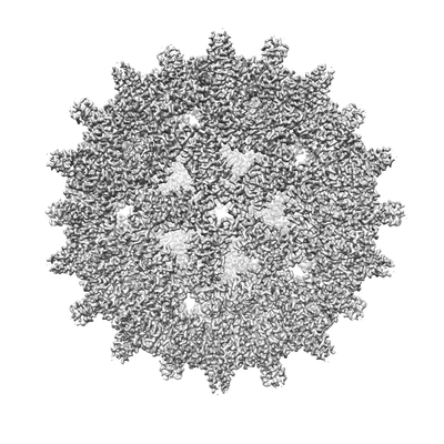
 Downloads & links
 emd_20669.png
 http://ftp.pdbj.org/pub/emdb/structures/EMD-20669


 Z (Sec.)
 Y (Row.)
 X (Col.)


 Sample components
 Processing
 Electron microscopy
 FIELD EMISSION GUN