 Movie
Movie Controller


+ Open data

- Basic information
| Entry | Database: PDB / ID: 1o7a | |||||||||
|---|---|---|---|---|---|---|---|---|---|---|
| Title | Human beta-Hexosaminidase B | |||||||||
 Components Components | BETA-HEXOSAMINIDASE BETA CHAIN | |||||||||
 Keywords Keywords | HYDROLASE / GLYCOSYL HYDROLASE / HEXOSAMINIDASE / LYSOSOMAL / SPHINGOLIPID DEGRADATION / SANDHOFF DISEASE / BA8-BARREL / GLYCOSIDASE | |||||||||
| Function / homology |  Function and homology information Function and homology informationbeta-N-acetylhexosaminidase complex / Defective HEXB causes GM2G2 (Hyaluronan metabolism) / chondroitin sulfate proteoglycan catabolic process / dermatan sulfate proteoglycan catabolic process / Keratan sulfate degradation / CS/DS degradation / Hyaluronan degradation / penetration of zona pellucida / male courtship behavior / N-acetylglucosamine metabolic process ...beta-N-acetylhexosaminidase complex / Defective HEXB causes GM2G2 (Hyaluronan metabolism) / chondroitin sulfate proteoglycan catabolic process / dermatan sulfate proteoglycan catabolic process / Keratan sulfate degradation / CS/DS degradation / Hyaluronan degradation / penetration of zona pellucida / male courtship behavior / N-acetylglucosamine metabolic process / cortical granule / glycosaminoglycan metabolic process / acetylglucosaminyltransferase activity / astrocyte cell migration / beta-N-acetylhexosaminidase activity / N-glycan processing / hyaluronan catabolic process / beta-N-acetylhexosaminidase / phospholipid biosynthetic process / Glycosphingolipid catabolism / lipid storage / ganglioside catabolic process / oligosaccharide catabolic process / azurophil granule / lysosome organization / neuromuscular process controlling balance / oogenesis / single fertilization / myelination / acrosomal vesicle / lysosomal lumen / beta-N-acetylglucosaminidase activity / skeletal system development / locomotory behavior / sensory perception of sound / neuron cellular homeostasis / intracellular calcium ion homeostasis / azurophil granule lumen / regulation of cell shape / carbohydrate binding / lysosome / Neutrophil degranulation / protein-containing complex binding / positive regulation of transcription by RNA polymerase II / extracellular exosome / extracellular region / identical protein binding / membrane Similarity search - Function | |||||||||
| Biological species |  HOMO SAPIENS (human) HOMO SAPIENS (human) | |||||||||
| Method |  X-RAY DIFFRACTION / SYNCHROTRON / MIRAS / Resolution: 2.25 Å X-RAY DIFFRACTION / SYNCHROTRON / MIRAS / Resolution: 2.25 Å | |||||||||
 Authors Authors | Maier, T. / Strater, N. / Schuette, C. / Klingenstein, R. / Sandhoff, K. / Saenger, W. | |||||||||
 Citation Citation | Journal: J.Mol.Biol. / Year: 2003 Title: The X-Ray Crystal Structure of Human Beta-Hexosaminidase B Provides New Insights Into Sandhoff Disease Authors: Maier, T. / Strater, N. / Schuette, C. / Klingenstein, R. / Sandhoff, K. / Saenger, W. #1: Journal: Glycobiology / Year: 2001 Title: Complete Analysis of the Glycosylation and Disulfide Bond Pattern of Human Beta-Hexosaminidase B by Maldi-Ms Authors: Schuette, C.G. / Weissgerber, J. / Sandhoff, K. #2: Journal: Protein Expr.Purif. / Year: 1990 Title: Synthesis of a Human Lysosomal Enzyme, Beta-Hexosaminidase B, Using the Baculaovirus Expression System Authors: Boose, J.A. / Tifft, C.J. / Proia, R.L. / Myerowitz, R. | |||||||||
| History |
| |||||||||
| Remark 650 | HELIX DETERMINATION METHOD: AUTHOR PROVIDED. | |||||||||
| Remark 700 | SHEET DETERMINATION METHOD: AUTHOR PROVIDED. |
- Structure visualization
| Structure viewer | Molecule: MolmilJmol/JSmol |
|---|
- Downloads & links
-Download
| PDBx/mmCIF format | 1o7a.cif.gz | 636 KB | Display | PDBx/mmCIF format |
|---|---|---|---|---|
| PDB format | pdb1o7a.ent.gz | 526.6 KB | Display | PDB format |
| PDBx/mmJSON format | 1o7a.json.gz | Tree view | PDBx/mmJSON format | |
| Others |  Other downloads Other downloads |
-Validation report
| Summary document | 1o7a_validation.pdf.gz | 2.2 MB | Display | wwPDB validaton report |
|---|---|---|---|---|
| Full document | 1o7a_full_validation.pdf.gz | 2.3 MB | Display | |
| Data in XML | 1o7a_validation.xml.gz | 135 KB | Display | |
| Data in CIF | 1o7a_validation.cif.gz | 193.1 KB | Display | |
| Arichive directory | https://data.pdbj.org/pub/pdb/validation_reports/o7/1o7aftp://data.pdbj.org/pub/pdb/validation_reports/o7/1o7a | HTTPS FTP |
-Related structure data
| Related structure data | |
|---|---|
| Similar structure data |


-Links
 PDBj
PDBj
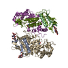
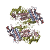
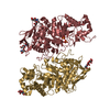
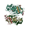
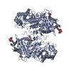
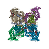
- Assembly
| Deposited unit | 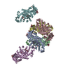
| ||||||||
|---|---|---|---|---|---|---|---|---|---|
| 1 | 
| ||||||||
| 2 | 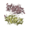
| ||||||||
| 3 | 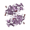
| ||||||||
| 4 | 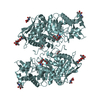
| ||||||||
| Unit cell |
|
-Components
-Protein , 1 types, 6 molecules ABCDEF
| #1: Protein | Mass: 58925.711 Da / Num. of mol.: 6 / Fragment: 42-556 Source method: isolated from a genetically manipulated source Details: RECOMBINANTLY EXPRESSED FRAGMENT COMPOSED OF RESIDUES 42-556, N-TERMINUS OF MATURE HUMAN ENZYME IS BETWEEN RESIDUE 48 - 50 Source: (gene. exp.) HOMO SAPIENS (human) / Plasmid: PACYM-1BETA / Cell line (production host): Sf21 / Production host:   SPODOPTERA FRUGIPERDA (fall armyworm) / References: UniProt: P07686, beta-N-acetylhexosaminidase SPODOPTERA FRUGIPERDA (fall armyworm) / References: UniProt: P07686, beta-N-acetylhexosaminidase |
|---|
-Sugars , 3 types, 18 molecules 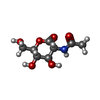

| #2: Polysaccharide | 2-acetamido-2-deoxy-beta-D-glucopyranose-(1-4)-2-acetamido-2-deoxy-beta-D-glucopyranose Source method: isolated from a genetically manipulated source #3: Sugar | ChemComp-GDL /  Type: D-saccharide / Mass: 219.192 Da / Num. of mol.: 6 / Source method: obtained synthetically / Formula: C8H13NO6 Type: D-saccharide / Mass: 219.192 Da / Num. of mol.: 6 / Source method: obtained synthetically / Formula: C8H13NO6#4: Sugar | ChemComp-NAG /  Type: D-saccharide, beta linking / Mass: 221.208 Da / Num. of mol.: 6 Type: D-saccharide, beta linking / Mass: 221.208 Da / Num. of mol.: 6Source method: isolated from a genetically manipulated source Formula: C8H15NO6 |
|---|
-Non-polymers , 2 types, 2341 molecules 

| #5: Chemical | ChemComp-EDO /  Mass: 62.068 Da / Num. of mol.: 15 / Source method: obtained synthetically / Formula: C2H6O2 Mass: 62.068 Da / Num. of mol.: 15 / Source method: obtained synthetically / Formula: C2H6O2#6: Water | ChemComp-HOH / | Mass: 18.015 Da / Num. of mol.: 2326 / Source method: isolated from a natural source / Formula: H2O |
|---|
-Details
| Compound details | BETA-HEXOSAMINIDASE IS RESPONSIBLE FOR THE DEGRADATION OF GM2 GANGLIOSIDES, AND A VARIETY OF OTHER ...BETA-HEXOSAMINI |
|---|---|
| Has protein modification | Y |
| Sequence details | EXPRESSED CONSTRUCT CONTAINS RESIDUES 42-TO 556 OF THE PRECURSOR PROTEIN |
-Experimental details
-Experiment
| Experiment | Method: X-RAY DIFFRACTION / Number of used crystals: 1 |
|---|
- Sample preparation
| Crystal | Density Matthews: 2.9 Å3/Da / Density % sol: 57 % | |||||||||||||||||||||||||||||||||||||||||||||||||
|---|---|---|---|---|---|---|---|---|---|---|---|---|---|---|---|---|---|---|---|---|---|---|---|---|---|---|---|---|---|---|---|---|---|---|---|---|---|---|---|---|---|---|---|---|---|---|---|---|---|---|
| Crystal grow | Method: vapor diffusion, hanging drop / pH: 5.6 Details: HANGING DROP VAPOUR DIFFUSION PROTEIN SOLUTION: 10 MG/ML HEXB IN 10MM SODIUM CITRATE, 100 MM NACL, PH6.0 RESERVOIR SOLUTION: 100 MM SODIUM CITRATE, PH 5.6, 16% (V/V) ETHYLENE GLYCOL, 10%(V/V) ...Details: HANGING DROP VAPOUR DIFFUSION PROTEIN SOLUTION: 10 MG/ML HEXB IN 10MM SODIUM CITRATE, 100 MM NACL, PH6.0 RESERVOIR SOLUTION: 100 MM SODIUM CITRATE, PH 5.6, 16% (V/V) ETHYLENE GLYCOL, 10%(V/V) POLYETHYLENE GLYCOL 8000 | |||||||||||||||||||||||||||||||||||||||||||||||||
| Crystal grow | *PLUS pH: 6 / Method: vapor diffusion, hanging drop | |||||||||||||||||||||||||||||||||||||||||||||||||
| Components of the solutions | *PLUS
|
-Data collection
| Diffraction | Mean temperature: 100 K |
|---|---|
| Diffraction source | Source: SYNCHROTRON / Site: ESRF  / Beamline: ID14-1 / Wavelength: 0.934 / Beamline: ID14-1 / Wavelength: 0.934 |
| Detector | Type: MARRESEARCH / Detector: CCD / Date: Mar 23, 2001 |
| Radiation | Protocol: SINGLE WAVELENGTH / Monochromatic (M) / Laue (L): M / Scattering type: x-ray |
| Radiation wavelength | Wavelength: 0.934 Å / Relative weight: 1 |
| Reflection | Resolution: 2.25→30 Å / Num. obs: 180325 / % possible obs: 99.9 % / Redundancy: 10.7 % / Biso Wilson estimate: 31.9 Å2 / Rmerge(I) obs: 0.065 / Net I/σ(I): 25.4 |
| Reflection shell | Resolution: 2.25→2.33 Å / Redundancy: 9.7 % / Rmerge(I) obs: 0.356 / Mean I/σ(I) obs: 5.5 / % possible all: 100 |
| Reflection | *PLUS Highest resolution: 2.25 Å / Lowest resolution: 30 Å / Num. measured all: 1930354 / Rmerge(I) obs: 0.065 |
| Reflection shell | *PLUS % possible obs: 100 % / Rmerge(I) obs: 0.356 / Mean I/σ(I) obs: 5.5 |
- Processing
| Software |
| ||||||||||||||||||||||||||||||||||||||||||||||||||||||||||||||||||||||||||||||||
|---|---|---|---|---|---|---|---|---|---|---|---|---|---|---|---|---|---|---|---|---|---|---|---|---|---|---|---|---|---|---|---|---|---|---|---|---|---|---|---|---|---|---|---|---|---|---|---|---|---|---|---|---|---|---|---|---|---|---|---|---|---|---|---|---|---|---|---|---|---|---|---|---|---|---|---|---|---|---|---|---|---|
| Refinement | Method to determine structure: MIRAS / Resolution: 2.25→28.12 Å / Rfactor Rfree error: 0.006 / Data cutoff high absF: 4883320.78 / Data cutoff low absF: 0 / Isotropic thermal model: RESTRAINED / Cross valid method: THROUGHOUT / σ(F): 0
| ||||||||||||||||||||||||||||||||||||||||||||||||||||||||||||||||||||||||||||||||
| Solvent computation | Solvent model: FLAT MODEL / Bsol: 56.6122 Å2 / ksol: 0.340616 e/Å3 | ||||||||||||||||||||||||||||||||||||||||||||||||||||||||||||||||||||||||||||||||
| Displacement parameters | Biso mean: 41.6 Å2
| ||||||||||||||||||||||||||||||||||||||||||||||||||||||||||||||||||||||||||||||||
| Refine analyze |
| ||||||||||||||||||||||||||||||||||||||||||||||||||||||||||||||||||||||||||||||||
| Refinement step | Cycle: LAST / Resolution: 2.25→28.12 Å
| ||||||||||||||||||||||||||||||||||||||||||||||||||||||||||||||||||||||||||||||||
| Refine LS restraints |
| ||||||||||||||||||||||||||||||||||||||||||||||||||||||||||||||||||||||||||||||||
| LS refinement shell | Resolution: 2.25→2.33 Å / Rfactor Rfree error: 0.023 / Total num. of bins used: 10
| ||||||||||||||||||||||||||||||||||||||||||||||||||||||||||||||||||||||||||||||||
| Xplor file |
| ||||||||||||||||||||||||||||||||||||||||||||||||||||||||||||||||||||||||||||||||
| Refine LS restraints | *PLUS
|
