 ムービー
ムービー コントローラー


+ データを開く

- 基本情報
基本情報
| 登録情報 | データベース: PDB / ID: 1o7a | |||||||||
|---|---|---|---|---|---|---|---|---|---|---|
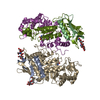
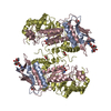
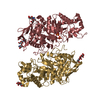
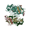
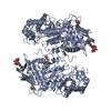
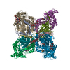
| タイトル | Human beta-Hexosaminidase B | |||||||||
 要素 要素 | BETA-HEXOSAMINIDASE BETA CHAIN | |||||||||
 キーワード キーワード | HYDROLASE / GLYCOSYL HYDROLASE / HEXOSAMINIDASE / LYSOSOMAL / SPHINGOLIPID DEGRADATION / SANDHOFF DISEASE / BA8-BARREL / GLYCOSIDASE | |||||||||
| 機能・相同性 |  機能・相同性情報 機能・相同性情報penetration of zona pellucida / beta-N-acetylhexosaminidase complex / Defective HEXB causes GM2G2 (Hyaluronan metabolism) / chondroitin sulfate proteoglycan catabolic process / dermatan sulfate proteoglycan catabolic process / Keratan sulfate degradation / CS/DS degradation / Hyaluronan degradation / male courtship behavior / N-acetylglucosamine metabolic process ...penetration of zona pellucida / beta-N-acetylhexosaminidase complex / Defective HEXB causes GM2G2 (Hyaluronan metabolism) / chondroitin sulfate proteoglycan catabolic process / dermatan sulfate proteoglycan catabolic process / Keratan sulfate degradation / CS/DS degradation / Hyaluronan degradation / male courtship behavior / N-acetylglucosamine metabolic process / glycosaminoglycan metabolic process / acetylglucosaminyltransferase activity / cortical granule / astrocyte cell migration / beta-N-acetylhexosaminidase activity / lipid storage / beta-N-acetylhexosaminidase / N-glycan processing / hyaluronan catabolic process / Glycosphingolipid catabolism / phospholipid biosynthetic process / ganglioside catabolic process / neuromuscular process controlling balance / oligosaccharide catabolic process / lysosome organization / azurophil granule / single fertilization / oogenesis / myelination / lysosomal lumen / acrosomal vesicle / beta-N-acetylglucosaminidase activity / skeletal system development / locomotory behavior / neuron cellular homeostasis / sensory perception of sound / intracellular calcium ion homeostasis / azurophil granule lumen / regulation of cell shape / carbohydrate binding / lysosome / Neutrophil degranulation / protein-containing complex binding / positive regulation of transcription by RNA polymerase II / extracellular exosome / extracellular region / membrane / identical protein binding 類似検索 - 分子機能 | |||||||||
| 生物種 |  HOMO SAPIENS (ヒト) HOMO SAPIENS (ヒト) | |||||||||
| 手法 |  X線回折 / シンクロトロン / 多重同系置換・異常分散 / 解像度: 2.25 Å X線回折 / シンクロトロン / 多重同系置換・異常分散 / 解像度: 2.25 Å | |||||||||
 データ登録者 データ登録者 | Maier, T. / Strater, N. / Schuette, C. / Klingenstein, R. / Sandhoff, K. / Saenger, W. | |||||||||
 引用 引用 | ジャーナル: J.Mol.Biol. / 年: 2003 タイトル: The X-Ray Crystal Structure of Human Beta-Hexosaminidase B Provides New Insights Into Sandhoff Disease 著者: Maier, T. / Strater, N. / Schuette, C. / Klingenstein, R. / Sandhoff, K. / Saenger, W. #1: ジャーナル: Glycobiology / 年: 2001 タイトル: Complete Analysis of the Glycosylation and Disulfide Bond Pattern of Human Beta-Hexosaminidase B by Maldi-Ms 著者: Schuette, C.G. / Weissgerber, J. / Sandhoff, K. #2: ジャーナル: Protein Expr.Purif. / 年: 1990 タイトル: Synthesis of a Human Lysosomal Enzyme, Beta-Hexosaminidase B, Using the Baculaovirus Expression System 著者: Boose, J.A. / Tifft, C.J. / Proia, R.L. / Myerowitz, R. | |||||||||
| 履歴 |
| |||||||||
| Remark 650 | HELIX DETERMINATION METHOD: AUTHOR PROVIDED. | |||||||||
| Remark 700 | SHEET DETERMINATION METHOD: AUTHOR PROVIDED. |
- 構造の表示
構造の表示
| 構造ビューア | 分子: MolmilJmol/JSmol |
|---|
- ダウンロードとリンク
-ダウンロード
| PDBx/mmCIF形式 | 1o7a.cif.gz | 636 KB | 表示 | PDBx/mmCIF形式 |
|---|---|---|---|---|
| PDB形式 | pdb1o7a.ent.gz | 526.6 KB | 表示 | PDB形式 |
| PDBx/mmJSON形式 | 1o7a.json.gz | ツリー表示 | PDBx/mmJSON形式 | |
| その他 |  その他のダウンロード その他のダウンロード |
-検証レポート
| アーカイブディレクトリ | https://data.pdbj.org/pub/pdb/validation_reports/o7/1o7aftp://data.pdbj.org/pub/pdb/validation_reports/o7/1o7a | HTTPS FTP |
|---|
-関連構造データ
| 関連構造データ | |
|---|---|
| 類似構造データ |


-リンク
 PDBj
PDBj
- 集合体
集合体
| 登録構造単位 | 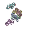
| ||||||||
|---|---|---|---|---|---|---|---|---|---|
| 1 | 
| ||||||||
| 2 | 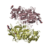
| ||||||||
| 3 | 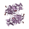
| ||||||||
| 4 | 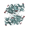
| ||||||||
| 単位格子 |
|
-要素
-タンパク質 , 1種, 6分子 ABCDEF
| #1: タンパク質 | 分子量: 58925.711 Da / 分子数: 6 / 断片: 42-556 / 由来タイプ: 組換発現 詳細: RECOMBINANTLY EXPRESSED FRAGMENT COMPOSED OF RESIDUES 42-556, N-TERMINUS OF MATURE HUMAN ENZYME IS BETWEEN RESIDUE 48 - 50 由来: (組換発現) HOMO SAPIENS (ヒト) / プラスミド: PACYM-1BETA / 細胞株 (発現宿主): Sf21発現宿主:   SPODOPTERA FRUGIPERDA (ツマジロクサヨトウ) SPODOPTERA FRUGIPERDA (ツマジロクサヨトウ)参照: UniProt: P07686, beta-N-acetylhexosaminidase |
|---|
-糖 , 3種, 18分子 

| #2: 多糖 | 2-acetamido-2-deoxy-beta-D-glucopyranose-(1-4)-2-acetamido-2-deoxy-beta-D-glucopyranose #3: 糖 | ChemComp-GDL /  タイプ: D-saccharide / 分子量: 219.192 Da / 分子数: 6 / 由来タイプ: 合成 / 式: C8H13NO6 タイプ: D-saccharide / 分子量: 219.192 Da / 分子数: 6 / 由来タイプ: 合成 / 式: C8H13NO6#4: 糖 | ChemComp-NAG /  タイプ: D-saccharide, beta linking / 分子量: 221.208 Da / 分子数: 6 / 由来タイプ: 組換発現 / 式: C8H15NO6 タイプ: D-saccharide, beta linking / 分子量: 221.208 Da / 分子数: 6 / 由来タイプ: 組換発現 / 式: C8H15NO6 |
|---|
-非ポリマー , 2種, 2341分子 

| #5: 化合物 | ChemComp-EDO /  分子量: 62.068 Da / 分子数: 15 / 由来タイプ: 合成 / 式: C2H6O2 分子量: 62.068 Da / 分子数: 15 / 由来タイプ: 合成 / 式: C2H6O2#6: 水 | ChemComp-HOH / | 分子量: 18.015 Da / 分子数: 2326 / 由来タイプ: 天然 / 式: H2O |
|---|
-詳細
| 構成要素の詳細 | BETA-HEXOSAMINIDASE IS RESPONSIBLE FOR THE DEGRADATION OF GM2 GANGLIOSIDES, AND A VARIETY OF OTHER ...BETA-HEXOSAMINI |
|---|---|
| Has protein modification | Y |
| 配列の詳細 | EXPRESSED CONSTRUCT CONTAINS RESIDUES 42-TO 556 OF THE PRECURSOR PROTEIN |
-実験情報
-実験
| 実験 | 手法: X線回折 / 使用した結晶の数: 1 |
|---|
- 試料調製
試料調製
| 結晶 | マシュー密度: 2.9 Å3/Da / 溶媒含有率: 57 % | |||||||||||||||||||||||||||||||||||||||||||||||||
|---|---|---|---|---|---|---|---|---|---|---|---|---|---|---|---|---|---|---|---|---|---|---|---|---|---|---|---|---|---|---|---|---|---|---|---|---|---|---|---|---|---|---|---|---|---|---|---|---|---|---|
| 結晶化 | 手法: 蒸気拡散法, ハンギングドロップ法 / pH: 5.6 詳細: HANGING DROP VAPOUR DIFFUSION PROTEIN SOLUTION: 10 MG/ML HEXB IN 10MM SODIUM CITRATE, 100 MM NACL, PH6.0 RESERVOIR SOLUTION: 100 MM SODIUM CITRATE, PH 5.6, 16% (V/V) ETHYLENE GLYCOL, 10%(V/V) ...詳細: HANGING DROP VAPOUR DIFFUSION PROTEIN SOLUTION: 10 MG/ML HEXB IN 10MM SODIUM CITRATE, 100 MM NACL, PH6.0 RESERVOIR SOLUTION: 100 MM SODIUM CITRATE, PH 5.6, 16% (V/V) ETHYLENE GLYCOL, 10%(V/V) POLYETHYLENE GLYCOL 8000 | |||||||||||||||||||||||||||||||||||||||||||||||||
| 結晶化 | *PLUS pH: 6 / 手法: 蒸気拡散法, ハンギングドロップ法 | |||||||||||||||||||||||||||||||||||||||||||||||||
| 溶液の組成 | *PLUS
|
-データ収集
| 回折 | 平均測定温度: 100 K |
|---|---|
| 放射光源 | 由来: シンクロトロン / サイト: ESRF  / ビームライン: ID14-1 / 波長: 0.934 / ビームライン: ID14-1 / 波長: 0.934 |
| 検出器 | タイプ: MARRESEARCH / 検出器: CCD / 日付: 2001年3月23日 |
| 放射 | プロトコル: SINGLE WAVELENGTH / 単色(M)・ラウエ(L): M / 散乱光タイプ: x-ray |
| 放射波長 | 波長: 0.934 Å / 相対比: 1 |
| 反射 | 解像度: 2.25→30 Å / Num. obs: 180325 / % possible obs: 99.9 % / 冗長度: 10.7 % / Biso Wilson estimate: 31.9 Å2 / Rmerge(I) obs: 0.065 / Net I/σ(I): 25.4 |
| 反射 シェル | 解像度: 2.25→2.33 Å / 冗長度: 9.7 % / Rmerge(I) obs: 0.356 / Mean I/σ(I) obs: 5.5 / % possible all: 100 |
| 反射 | *PLUS 最高解像度: 2.25 Å / 最低解像度: 30 Å / Num. measured all: 1930354 / Rmerge(I) obs: 0.065 |
| 反射 シェル | *PLUS % possible obs: 100 % / Rmerge(I) obs: 0.356 / Mean I/σ(I) obs: 5.5 |
- 解析
解析
| ソフトウェア |
| ||||||||||||||||||||||||||||||||||||||||||||||||||||||||||||||||||||||||||||||||
|---|---|---|---|---|---|---|---|---|---|---|---|---|---|---|---|---|---|---|---|---|---|---|---|---|---|---|---|---|---|---|---|---|---|---|---|---|---|---|---|---|---|---|---|---|---|---|---|---|---|---|---|---|---|---|---|---|---|---|---|---|---|---|---|---|---|---|---|---|---|---|---|---|---|---|---|---|---|---|---|---|---|
| 精密化 | 構造決定の手法: 多重同系置換・異常分散 / 解像度: 2.25→28.12 Å / Rfactor Rfree error: 0.006 / Data cutoff high absF: 4883320.78 / Data cutoff low absF: 0 / Isotropic thermal model: RESTRAINED / 交差検証法: THROUGHOUT / σ(F): 0
| ||||||||||||||||||||||||||||||||||||||||||||||||||||||||||||||||||||||||||||||||
| 溶媒の処理 | 溶媒モデル: FLAT MODEL / Bsol: 56.6122 Å2 / ksol: 0.340616 e/Å3 | ||||||||||||||||||||||||||||||||||||||||||||||||||||||||||||||||||||||||||||||||
| 原子変位パラメータ | Biso mean: 41.6 Å2
| ||||||||||||||||||||||||||||||||||||||||||||||||||||||||||||||||||||||||||||||||
| Refine analyze |
| ||||||||||||||||||||||||||||||||||||||||||||||||||||||||||||||||||||||||||||||||
| 精密化ステップ | サイクル: LAST / 解像度: 2.25→28.12 Å
| ||||||||||||||||||||||||||||||||||||||||||||||||||||||||||||||||||||||||||||||||
| 拘束条件 |
| ||||||||||||||||||||||||||||||||||||||||||||||||||||||||||||||||||||||||||||||||
| LS精密化 シェル | 解像度: 2.25→2.33 Å / Rfactor Rfree error: 0.023 / Total num. of bins used: 10
| ||||||||||||||||||||||||||||||||||||||||||||||||||||||||||||||||||||||||||||||||
| Xplor file |
| ||||||||||||||||||||||||||||||||||||||||||||||||||||||||||||||||||||||||||||||||
| 拘束条件 | *PLUS
|
