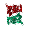
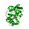
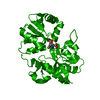
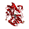
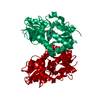
登録情報 データベース : PDB / ID : 1mqiタイトル Crystal Structure of the GluR2 Ligand Binding Core (S1S2J) in Complex with Fluoro-Willardiine at 1.35 Angstroms Resolution glutamate receptor 2 キーワード / / / / / / / 機能・相同性 分子機能 ドメイン・相同性 構成要素
/ / / / / / / / / / / / / / / / / / / / / / / / / / / / / / / / / / / / / / / / / / / / / / / / / / / / / / / / / / / / / / / / / / / / / / / / / / / / / / / / / / / / / / / / / / / / / / / / / / / / / / / / / / 生物種 Rattus norvegicus (ドブネズミ)手法 / / / 解像度 : 1.35 Å データ登録者 Jin, R. / Banke, T.G. / Mayer, M.L. / Traynelis, S.F. / Gouaux, E. 履歴 登録 2002年9月16日 登録サイト / 処理サイト 改定 1.0 2003年8月5日 Provider / タイプ 改定 1.1 2008年4月28日 Group 改定 1.2 2011年7月13日 Group 改定 1.3 2017年8月2日 Group / Source and taxonomy / カテゴリ / software
すべて表示 表示を減らす
 ムービー
ムービー コントローラー


 データを開く
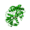
 基本情報
基本情報 要素
要素 キーワード
キーワード 機能・相同性情報
 X線回折 /
  データ登録者
 引用
引用 構造の表示
構造の表示 ダウンロードとリンク
 その他のダウンロード


 PDBj
PDBj


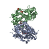
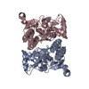
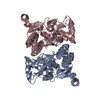
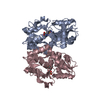
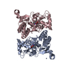
 集合体
集合体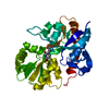

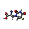
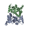
 分子量: 217.155 Da / 分子数: 1 / 由来タイプ: 合成 / 式: C7H8FN3O4
 分子量: 18.015 Da / 分子数: 382 / 由来タイプ: 天然 / 式: H2O
 試料調製
試料調製 / ビームライン: X4A / 波長: 0.9746 Å
 解析
解析