 Movie
Movie Controller

[English] 日本語
 Yorodumi
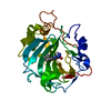
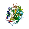
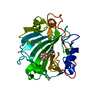
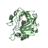
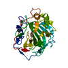
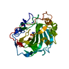
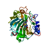
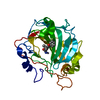
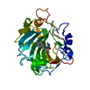
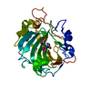
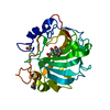
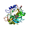
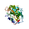
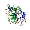
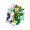
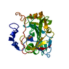
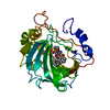
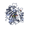
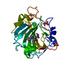
- PDB-1hcb: ENZYME-SUBSTRATE INTERACTIONS: STRUCTURE OF HUMAN CARBONIC ANHYDR... -
+ Open data

- Basic information
| Entry | Database: PDB / ID: 1hcb | ||||||
|---|---|---|---|---|---|---|---|
| Title | ENZYME-SUBSTRATE INTERACTIONS: STRUCTURE OF HUMAN CARBONIC ANHYDRASE I COMPLEXED WITH BICARBONATE | ||||||
 Components Components | CARBONIC ANHYDRASE I | ||||||
 Keywords Keywords | LYASE(OXO-ACID) | ||||||
| Function / homology |  Function and homology information Function and homology informationhydro-lyase activity / response to fructose / cyanamide hydratase / cyanamide hydratase activity / arylesterase activity / Gene and protein expression by JAK-STAT signaling after Interleukin-12 stimulation / Reversible hydration of carbon dioxide / carbonic anhydrase / carbonate dehydratase activity / Erythrocytes take up oxygen and release carbon dioxide ...hydro-lyase activity / response to fructose / cyanamide hydratase / cyanamide hydratase activity / arylesterase activity / Gene and protein expression by JAK-STAT signaling after Interleukin-12 stimulation / Reversible hydration of carbon dioxide / carbonic anhydrase / carbonate dehydratase activity / Erythrocytes take up oxygen and release carbon dioxide / Erythrocytes take up carbon dioxide and release oxygen / extracellular exosome / zinc ion binding / cytoplasm / cytosol Similarity search - Function | ||||||
| Biological species |  Homo sapiens (human) Homo sapiens (human) | ||||||
| Method |  X-RAY DIFFRACTION / Resolution: 1.6 Å X-RAY DIFFRACTION / Resolution: 1.6 Å | ||||||
 Authors Authors | Kumar, V. / Kannan, K.K. | ||||||
 Citation Citation | Journal: J.Mol.Biol. / Year: 1994 Title: Enzyme-substrate interactions. Structure of human carbonic anhydrase I complexed with bicarbonate. Authors: Kumar, V. / Kannan, K.K. #1: Journal: Acta Crystallogr.,Sect.A / Year: 1993Title: Structure of Human Carbonic Anhydrase I Complexed with Gold Cyanide Inhibitor: Inhibition Mechanism Authors: Kumar, V. / Kannan, K.K. #2: Journal: Acta Crystallogr.,Sect.A / Year: 1987Title: Human Carbonic Anhydrase I-Iodide Complex: Structure and Inhibition Mechanism Authors: Kumar, V. / Satyamurthy, P. / Kannan, K.K. | ||||||
| History |
| ||||||
| Remark 700 | SHEET THIS STRUCTURE CONTAINS A BIFURCATED SHEET. THIS IS REPRESENTED BY PRESENTING TWO SHEETS ON ...SHEET THIS STRUCTURE CONTAINS A BIFURCATED SHEET. THIS IS REPRESENTED BY PRESENTING TWO SHEETS ON *SHEET* RECORDS BELOW. STRAND 5 IS DIFFERENT IN SHEETS S1A AND S1B. |
- Structure visualization
| Structure viewer | Molecule: MolmilJmol/JSmol |
|---|
- Downloads & links
-Download
| PDBx/mmCIF format | 1hcb.cif.gz | 69.5 KB | Display | PDBx/mmCIF format |
|---|---|---|---|---|
| PDB format | pdb1hcb.ent.gz | 51.1 KB | Display | PDB format |
| PDBx/mmJSON format | 1hcb.json.gz | Tree view | PDBx/mmJSON format | |
| Others |  Other downloads Other downloads |
-Validation report
| Arichive directory | https://data.pdbj.org/pub/pdb/validation_reports/hc/1hcbftp://data.pdbj.org/pub/pdb/validation_reports/hc/1hcb | HTTPS FTP |
|---|
-Related structure data
| Similar structure data |
|---|
-Links
 PDBj
PDBj


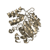
- Assembly
| Deposited unit | 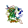
| ||||||||
|---|---|---|---|---|---|---|---|---|---|
| 1 |
| ||||||||
| Unit cell |
| ||||||||
| Atom site foot note | 1: CIS PROLINE - PRO 30 / 2: CIS PROLINE - PRO 202 |
-Components
| #1: Protein | Mass: 28774.988 Da / Num. of mol.: 1 Source method: isolated from a genetically manipulated source Source: (gene. exp.) Homo sapiens (human) / References: UniProt: P00915, carbonic anhydrase |
|---|---|
| #2: Chemical | ChemComp-ZN /   Mass: 65.409 Da / Num. of mol.: 1 / Source method: obtained synthetically / Formula: Zn Mass: 65.409 Da / Num. of mol.: 1 / Source method: obtained synthetically / Formula: Zn |
| #3: Chemical | ChemComp-BCT /   Mass: 61.017 Da / Num. of mol.: 1 / Source method: obtained synthetically / Formula: CHO3 / Comment: pH buffer*YM Mass: 61.017 Da / Num. of mol.: 1 / Source method: obtained synthetically / Formula: CHO3 / Comment: pH buffer*YM |
| #4: Water | ChemComp-HOH /  Mass: 18.015 Da / Num. of mol.: 254 / Source method: isolated from a natural source / Formula: H2O Mass: 18.015 Da / Num. of mol.: 254 / Source method: isolated from a natural source / Formula: H2O |
| Nonpolymer details | ATOM O HOH 520 (PARTIAL OCCUPANCY) MAKES SHORT CONTACT WITH 522 HCO GROUP AND MAY BE THE PARTIALLY ...ATOM O HOH 520 (PARTIAL OCCUPANCY) MAKES SHORT CONTACT WITH 522 HCO GROUP AND MAY BE THE PARTIALLY OVERLAPPIN |
| Sequence details | ON THE BASIS OF THE PRESENT COMPLEX STRUCTURE THE FOLLOWING SEQUENCE MODIFICATION IN THE NATIVE ...ON THE BASIS OF THE PRESENT COMPLEX STRUCTURE THE FOLLOWING SEQUENCE MODIFICATI |
-Experimental details
-Experiment
| Experiment | Method: X-RAY DIFFRACTION |
|---|
- Sample preparation
| Crystal | Density Matthews: 2.02 Å3/Da / Density % sol: 39.14 % |
|---|---|
| Crystal grow | *PLUS Method: unknown |
-Data collection
| Radiation | Scattering type: x-ray |
|---|---|
| Radiation wavelength | Relative weight: 1 |
| Reflection | *PLUS Highest resolution: 1.6 Å / Num. obs: 24976 / % possible obs: 79.1 % / Rmerge(I) obs: 0.0817 |
- Processing
| Software | Name: PROLSQ / Classification: refinement | |||||||||||||||||||||||||||||||||||||||||||||||||||||||||||||||
|---|---|---|---|---|---|---|---|---|---|---|---|---|---|---|---|---|---|---|---|---|---|---|---|---|---|---|---|---|---|---|---|---|---|---|---|---|---|---|---|---|---|---|---|---|---|---|---|---|---|---|---|---|---|---|---|---|---|---|---|---|---|---|---|---|
| Refinement | Resolution: 1.6→10 Å Details: THE SIDE CHAINS OF RESIDUES ASP 4, ASP 9, LYS 10, LEU 19 AND LYS149 SHOW VERY POOR ELECTRON DENSITY IN THE FOURIER MAPS.
| |||||||||||||||||||||||||||||||||||||||||||||||||||||||||||||||
| Refinement step | Cycle: LAST / Resolution: 1.6→10 Å
| |||||||||||||||||||||||||||||||||||||||||||||||||||||||||||||||
| Refine LS restraints |
| |||||||||||||||||||||||||||||||||||||||||||||||||||||||||||||||
| Software | *PLUS Name: PROLSQ / Classification: refinement | |||||||||||||||||||||||||||||||||||||||||||||||||||||||||||||||
| Refinement | *PLUS Highest resolution: 1.6 Å / Lowest resolution: 10 Å / Num. reflection obs: 24976 / Rfactor obs: 0.177 | |||||||||||||||||||||||||||||||||||||||||||||||||||||||||||||||
| Solvent computation | *PLUS | |||||||||||||||||||||||||||||||||||||||||||||||||||||||||||||||
| Displacement parameters | *PLUS |
