 Movie
Movie Controller

[English] 日本語
 Yorodumi
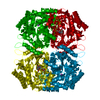
- PDB-1d7u: Crystal structure of the complex of 2,2-dialkylglycine decarboxyl... -
+ Open data

- Basic information
| Entry | Database: PDB / ID: 1d7u | ||||||
|---|---|---|---|---|---|---|---|
| Title | Crystal structure of the complex of 2,2-dialkylglycine decarboxylase with LCS | ||||||
 Components Components | PROTEIN (2,2-DIALKYLGLYCINE DECARBOXYLASE (PYRUVATE)) | ||||||
 Keywords Keywords | LYASE / ENZYME COMPLEXES / CATALYTIC MECHANISM / DECARBOXYLATION INHIBITOR | ||||||
| Function / homology |  Function and homology information Function and homology information2,2-dialkylglycine decarboxylase (pyruvate) / 2,2-dialkylglycine decarboxylase (pyruvate) activity / transaminase activity / pyridoxal phosphate binding Similarity search - Function | ||||||
| Biological species |  Burkholderia cepacia (bacteria) Burkholderia cepacia (bacteria) | ||||||
| Method |  X-RAY DIFFRACTION / SYNCHROTRON / MOLECULAR REPLACEMENT / Resolution: 1.95 Å X-RAY DIFFRACTION / SYNCHROTRON / MOLECULAR REPLACEMENT / Resolution: 1.95 Å | ||||||
 Authors Authors | Malashkevich, V.N. / Toney, M.D. / Strop, P. / Keller, J. / Jansonius, J.N. | ||||||
 Citation Citation | Journal: J.Mol.Biol. / Year: 1999 Title: Crystal structures of dialkylglycine decarboxylase inhibitor complexes. Authors: Malashkevich, V.N. / Strop, P. / Keller, J.W. / Jansonius, J.N. / Toney, M.D. #1: Journal: J.Mol.Biol. / Year: 1991Title: Crystallization and Preliminary X-Ray Diffraction Studies of Dialkylglycine Decarboxylase, a Decarboxylating Transaminase Authors: Toney, M.D. / Keller, J.W. / Pauptit, R.A. / Jaeger, J. / Wise, M.K. / Sauder, U. / Jansonius, J.N. #2: Journal: J.Biol.Chem. / Year: 1990Title: Pseudomonas cepacia 2,2-Dialkylglycine Decarboxylase. Sequence and Expression in Escherichia Coli of Structural and Repressor Genes Authors: Keller, J.W. / Baurick, K.B. / Rutt, G.C. / O'Malley, M.V. / Sonafrank, N.L. / Reynolds, R.A. / Ebbesson, L.O. / Vajdos, F.F. | ||||||
| History |
|
- Structure visualization
| Structure viewer | Molecule: MolmilJmol/JSmol |
|---|
- Downloads & links
-Download
| PDBx/mmCIF format | 1d7u.cif.gz | 99 KB | Display | PDBx/mmCIF format |
|---|---|---|---|---|
| PDB format | pdb1d7u.ent.gz | 73.8 KB | Display | PDB format |
| PDBx/mmJSON format | 1d7u.json.gz | Tree view | PDBx/mmJSON format | |
| Others |  Other downloads Other downloads |
-Validation report
| Arichive directory | https://data.pdbj.org/pub/pdb/validation_reports/d7/1d7uftp://data.pdbj.org/pub/pdb/validation_reports/d7/1d7u | HTTPS FTP |
|---|
-Related structure data
| Related structure data |  1d7rC  1d7sC  1d7vC  2dkbS C: citing same article ( S: Starting model for refinement |
|---|---|
| Similar structure data |


-Links
 PDBj
PDBj- Assembly
| Deposited unit | 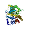
| ||||||||
|---|---|---|---|---|---|---|---|---|---|
| 1 | 
| ||||||||
| Unit cell |
|
-Components
| #1: Protein | Mass: 46495.320 Da / Num. of mol.: 1 Source method: isolated from a genetically manipulated source Source: (gene. exp.) Burkholderia cepacia (bacteria) / Plasmid: PKDHE19 / Production host: References: UniProt: P16932, 2,2-dialkylglycine decarboxylase (pyruvate) |
|---|---|
| #2: Chemical | ChemComp-NA /   Mass: 22.990 Da / Num. of mol.: 1 / Source method: obtained synthetically / Formula: Na Mass: 22.990 Da / Num. of mol.: 1 / Source method: obtained synthetically / Formula: Na |
| #3: Chemical | ChemComp-K /   Mass: 39.098 Da / Num. of mol.: 1 / Source method: obtained synthetically / Formula: K Mass: 39.098 Da / Num. of mol.: 1 / Source method: obtained synthetically / Formula: K |
| #4: Chemical | ChemComp-LCS / [  Mass: 331.219 Da / Num. of mol.: 1 / Source method: obtained synthetically / Formula: C11H14N3O7P Mass: 331.219 Da / Num. of mol.: 1 / Source method: obtained synthetically / Formula: C11H14N3O7P |
| #5: Water | ChemComp-HOH /  Mass: 18.015 Da / Num. of mol.: 148 / Source method: isolated from a natural source / Formula: H2O Mass: 18.015 Da / Num. of mol.: 148 / Source method: isolated from a natural source / Formula: H2O |
-Experimental details
-Experiment
| Experiment | Method: X-RAY DIFFRACTION / Number of used crystals: 1 |
|---|
- Sample preparation
| Crystal | Density Matthews: 3.15 Å3/Da / Density % sol: 61 % | ||||||||||||||||||||||||||||||||||||||||||
|---|---|---|---|---|---|---|---|---|---|---|---|---|---|---|---|---|---|---|---|---|---|---|---|---|---|---|---|---|---|---|---|---|---|---|---|---|---|---|---|---|---|---|---|
| Crystal grow | Temperature: 297 K / Method: vapor diffusion, hanging drop / pH: 7.5 Details: pH 7.50, VAPOR DIFFUSION, HANGING DROP, temperature 297K | ||||||||||||||||||||||||||||||||||||||||||
| Crystal | *PLUS | ||||||||||||||||||||||||||||||||||||||||||
| Crystal grow | *PLUS pH: 7.5 | ||||||||||||||||||||||||||||||||||||||||||
| Components of the solutions | *PLUS
|
-Data collection
| Diffraction | Mean temperature: 100 K |
|---|---|
| Diffraction source | Source: SYNCHROTRON / Site: MPG/DESY, HAMBURG  / Beamline: BW6 / Wavelength: 1.1 / Beamline: BW6 / Wavelength: 1.1 |
| Detector | Type: MARRESEARCH / Detector: IMAGE PLATE / Date: Jun 26, 1996 |
| Radiation | Protocol: SINGLE WAVELENGTH / Monochromatic (M) / Laue (L): M / Scattering type: x-ray |
| Radiation wavelength | Wavelength: 1.1 Å / Relative weight: 1 |
| Reflection | Resolution: 1.95→35 Å / Num. obs: 42043 / % possible obs: 95.6 % / Observed criterion σ(I): -3 / Redundancy: 3.9 % / Biso Wilson estimate: 27.04 Å2 / Rmerge(I) obs: 0.062 / Net I/σ(I): 9.8 |
| Reflection shell | Resolution: 1.95→2 Å / Redundancy: 2.5 % / Rmerge(I) obs: 0.398 / % possible all: 73.3 |
| Reflection | *PLUS Redundancy: 3.9 % |
- Processing
| Software |
| ||||||||||||
|---|---|---|---|---|---|---|---|---|---|---|---|---|---|
| Refinement | Method to determine structure: MOLECULAR REPLACEMENT Starting model: 2DKB Resolution: 1.95→10 Å / Isotropic thermal model: RESTRAINED / σ(F): 2 / Stereochemistry target values: Engh & Huber
| ||||||||||||
| Solvent computation | Solvent model: FLAT MODEL / Bsol: 164.5 Å2 / ksol: 0.67 e/Å3 | ||||||||||||
| Displacement parameters | Biso mean: 35.26 Å2 | ||||||||||||
| Refine analyze | Luzzati coordinate error obs: 0.22 Å / Luzzati d res low obs: 5 Å / Luzzati sigma a obs: 0.22 Å | ||||||||||||
| Refinement step | Cycle: LAST / Resolution: 1.95→10 Å
| ||||||||||||
| Refine LS restraints |
| ||||||||||||
| Software | *PLUS Name: TNT / Classification: refinement | ||||||||||||
| Refinement | *PLUS Rfactor obs: 0.193 | ||||||||||||
| Solvent computation | *PLUS | ||||||||||||
| Displacement parameters | *PLUS | ||||||||||||
| Refine LS restraints | *PLUS
|
