 Movie
Movie Controller

[English] 日本語
 Yorodumi
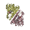
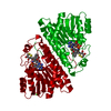
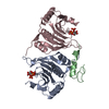
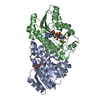
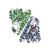
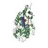
- PDB-1b16: ALCOHOL DEHYDROGENASE FROM DROSOPHILA LEBANONENSIS TERNARY COMPLE... -
+ Open data

- Basic information
| Entry | Database: PDB / ID: 1b16 | ||||||
|---|---|---|---|---|---|---|---|
| Title | ALCOHOL DEHYDROGENASE FROM DROSOPHILA LEBANONENSIS TERNARY COMPLEX WITH NAD-3-PENTANONE | ||||||
 Components Components | PROTEIN (ALCOHOL DEHYDROGENASE) | ||||||
 Keywords Keywords | OXIDOREDUCTASE / DETOXIFICATION / METABOLISM / ALCOHOL DEHYDROGENASE / DROSOPHILA LEBANONENSIS / SHORT-CHAIN DEHYDROGENASES/REDUCTASES / TERNARY COMPLEX / NAD-3- PENTANONE ADDUCT | ||||||
| Function / homology |  Function and homology information Function and homology informationalcohol metabolic process / alcohol dehydrogenase (NAD+) activity / alcohol dehydrogenase / identical protein binding / cytoplasm Similarity search - Function | ||||||
| Biological species |  Scaptodrosophila lebanonensis (fry) Scaptodrosophila lebanonensis (fry) | ||||||
| Method |  X-RAY DIFFRACTION / SYNCHROTRON / MOLECULAR REPLACEMENT / Resolution: 1.4 Å X-RAY DIFFRACTION / SYNCHROTRON / MOLECULAR REPLACEMENT / Resolution: 1.4 Å | ||||||
 Authors Authors | Benach, J. / Atrian, S. / Gonzalez-Duarte, R. / Ladenstein, R. | ||||||
 Citation Citation | Journal: J.Mol.Biol. / Year: 1999 Title: The catalytic reaction and inhibition mechanism of Drosophila alcohol dehydrogenase: observation of an enzyme-bound NAD-ketone adduct at 1.4 A resolution by X-ray crystallography. Authors: Benach, J. / Atrian, S. / Gonzalez-Duarte, R. / Ladenstein, R. #1: Journal: J.Mol.Biol. / Year: 1998Title: The Refined Crystal Structure of Drosophila Lebanonensis Alcohol Dehydrogenase at 1.9 A Resolution Authors: Benach, J. / Atrian, S. / Gonzalez-Duarte, R. / Ladenstein, R. | ||||||
| History |
|
- Structure visualization
| Structure viewer | Molecule: MolmilJmol/JSmol |
|---|
- Downloads & links
-Download
| PDBx/mmCIF format | 1b16.cif.gz | 123.6 KB | Display | PDBx/mmCIF format |
|---|---|---|---|---|
| PDB format | pdb1b16.ent.gz | 94.6 KB | Display | PDB format |
| PDBx/mmJSON format | 1b16.json.gz | Tree view | PDBx/mmJSON format | |
| Others |  Other downloads Other downloads |
-Validation report
| Summary document | 1b16_validation.pdf.gz | 529.8 KB | Display | wwPDB validaton report |
|---|---|---|---|---|
| Full document | 1b16_full_validation.pdf.gz | 538.3 KB | Display | |
| Data in XML | 1b16_validation.xml.gz | 13.5 KB | Display | |
| Data in CIF | 1b16_validation.cif.gz | 21.8 KB | Display | |
| Arichive directory | https://data.pdbj.org/pub/pdb/validation_reports/b1/1b16ftp://data.pdbj.org/pub/pdb/validation_reports/b1/1b16 | HTTPS FTP |
-Related structure data
| Related structure data |  1b14C  1b15C  1b2lC  1a4uS S: Starting model for refinement C: citing same article ( |
|---|---|
| Similar structure data |


-Links
 PDBj
PDBj


- Assembly
| Deposited unit | 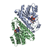
| ||||||||
|---|---|---|---|---|---|---|---|---|---|
| 1 |
| ||||||||
| Unit cell |
| ||||||||
| Noncrystallographic symmetry (NCS) | NCS oper: (Code: given Matrix: (-0.983274, -0.169734, 0.066045), Vector: |
-Components
| #1: Protein | Mass: 27823.973 Da / Num. of mol.: 2 / Source method: isolated from a natural source / Details: NAD-3-PENTANONE / Source: (natural) Scaptodrosophila lebanonensis (fry) / References: UniProt: P10807, alcohol dehydrogenase#2: Chemical | 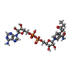  Mass: 747.542 Da / Num. of mol.: 2 / Source method: obtained synthetically / Formula: C26H35N7O15P2 Mass: 747.542 Da / Num. of mol.: 2 / Source method: obtained synthetically / Formula: C26H35N7O15P2#3: Water | ChemComp-HOH / |  Mass: 18.015 Da / Num. of mol.: 485 / Source method: isolated from a natural source / Formula: H2O Mass: 18.015 Da / Num. of mol.: 485 / Source method: isolated from a natural source / Formula: H2ONonpolymer details | NAD-3-PENTANONE ADDUCT | |
|---|
-Experimental details
-Experiment
| Experiment | Method: X-RAY DIFFRACTION / Number of used crystals: 1 |
|---|
- Sample preparation
| Crystal | Density Matthews: 2.15 Å3/Da / Density % sol: 45.5 % | ||||||||||||||||||||||||||||||||||||||||||||||||
|---|---|---|---|---|---|---|---|---|---|---|---|---|---|---|---|---|---|---|---|---|---|---|---|---|---|---|---|---|---|---|---|---|---|---|---|---|---|---|---|---|---|---|---|---|---|---|---|---|---|
| Crystal grow | pH: 7.5 Details: PROTEIN WAS CRYSTALLIZED FROM 28% PEG 2000, 0.2 M CACL2, 0.1 M TRIS-HCL, PH= 7.5, 1MM NAD+, 1% 3-PENTANONE, 4 C. | ||||||||||||||||||||||||||||||||||||||||||||||||
| Crystal grow | *PLUS Temperature: 277 K / pH: 8.6 / Method: vapor diffusion, sitting drop | ||||||||||||||||||||||||||||||||||||||||||||||||
| Components of the solutions | *PLUS
|
-Data collection
| Diffraction | Mean temperature: 100 K |
|---|---|
| Diffraction source | Source: SYNCHROTRON / Site: EMBL/DESY, HAMBURG  / Beamline: X31 / Wavelength: 0.9511 / Beamline: X31 / Wavelength: 0.9511 |
| Detector | Type: MARRESEARCH / Detector: IMAGE PLATE / Date: Nov 15, 1997 / Details: MIRROR |
| Radiation | Monochromator: SI(111) / Protocol: SINGLE WAVELENGTH / Monochromatic (M) / Laue (L): M / Scattering type: x-ray |
| Radiation wavelength | Wavelength: 0.9511 Å / Relative weight: 1 |
| Reflection | Resolution: 1.32→15 Å / Num. obs: 103695 / % possible obs: 94 % / Observed criterion σ(I): -3 / Redundancy: 3 % / Biso Wilson estimate: 13.2 Å2 / Rmerge(I) obs: 0.06 / Net I/σ(I): 3 |
| Reflection shell | Resolution: 1.32→1.43 Å / Redundancy: 3 % / Rmerge(I) obs: 0.4 / Mean I/σ(I) obs: 3 / % possible all: 94 |
| Reflection | *PLUS Num. measured all: 262892 |
| Reflection shell | *PLUS % possible obs: 94 % |
- Processing
| Software |
| ||||||||||||||||||||||||||||
|---|---|---|---|---|---|---|---|---|---|---|---|---|---|---|---|---|---|---|---|---|---|---|---|---|---|---|---|---|---|
| Refinement | Method to determine structure: MOLECULAR REPLACEMENT Starting model: 1A4U Resolution: 1.4→8 Å / Cross valid method: THROUGHOUT / σ(F): 0 / ESU R: 0.15 / ESU R Free: 0.17
| ||||||||||||||||||||||||||||
| Displacement parameters | Biso mean: 13.2 Å2 | ||||||||||||||||||||||||||||
| Refinement step | Cycle: LAST / Resolution: 1.4→8 Å
| ||||||||||||||||||||||||||||
| Software | *PLUS Name: REFMAC / Classification: refinement | ||||||||||||||||||||||||||||
| Refine LS restraints | *PLUS
|
