- EMDB-16968: Small subunit of yeast mitochondrial ribosome. -
+
Open data
ID or keywords:
Loading...
-
Basic information
Entry
Database: EMDB / ID: EMD-16968
Title
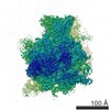
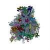
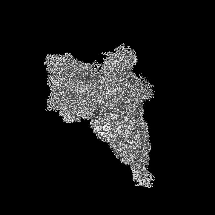
Small subunit of yeast mitochondrial ribosome.
Map data
Oversampled composite map of local-mask refined maps with sharpening and local resolution filtering
Sample
Complex: Small subunit of mitochondrial ribosome
Protein or peptide: x 32 types
RNA: x 1 types
Protein or peptide: x 1 types
Ligand: x 5 types
Keywords
mitochondria / RIBOSOME
Function / homology
Function and homology information
Branched-chain amino acid catabolism / 3-hydroxyisobutyryl-CoA hydrolase / 3-hydroxyisobutyryl-CoA hydrolase activity / mitochondrial translational initiation / valine catabolic process / Mitochondrial protein degradation / mitochondrial ribosome assembly / mitochondrial small ribosomal subunit / mitochondrial ribosome / sporulation resulting in formation of a cellular spore ...Branched-chain amino acid catabolism / 3-hydroxyisobutyryl-CoA hydrolase / 3-hydroxyisobutyryl-CoA hydrolase activity / mitochondrial translational initiation / valine catabolic process / Mitochondrial protein degradation / mitochondrial ribosome assembly / mitochondrial small ribosomal subunit / mitochondrial ribosome / sporulation resulting in formation of a cellular spore / mitochondrial translation / superoxide dismutase activity / mRNA processing / ribosomal small subunit biogenesis / small ribosomal subunit rRNA binding / peroxisome / ribosomal small subunit assembly / small ribosomal subunit / mitochondrial inner membrane / rRNA binding / ribosome / structural constituent of ribosome / translation / mRNA binding / GTP binding / mitochondrion / RNA binding / ATP binding / metal ion binding / cytoplasm Similarity search - Function
Ribosomal protein VAR1 / : / : / Mitochondrial ribosomal protein (VAR1) / Mitochondrial ribosomal protein MRP51, fungi / Mitochondrial ribosomal protein S25 / Ribosomal protein S24, mitochondrial / Ribosomal protein S23, mitochondrial, fungi / Mitochondrial ribosomal protein subunit / Mitochondrial ribosomal protein S25 ...Ribosomal protein VAR1 / : / : / Mitochondrial ribosomal protein (VAR1) / Mitochondrial ribosomal protein MRP51, fungi / Mitochondrial ribosomal protein S25 / Ribosomal protein S24, mitochondrial / Ribosomal protein S23, mitochondrial, fungi / Mitochondrial ribosomal protein subunit / Mitochondrial ribosomal protein S25 / Ribosomal protein MRP10, mitochondrial / Ribosomal protein S35, mitochondrial / Eukaryotic mitochondrial regulator protein / Enoyl-CoA hydratase/isomerase domain / IGR protein motif / Enoyl-CoA hydratase/isomerase, HIBYL-CoA-H type / Enoyl-CoA hydratase/isomerase / Protein Fyv4 / IGR protein motif / IGR / Ribosomal protein S27/S33, mitochondrial / Ribosomal protein S24/S35, mitochondrial / Mitochondrial ribosomal subunit S27 / Ribosomal protein S24/S35, mitochondrial, conserved domain / Mitochondrial ribosomal subunit protein / Enoyl-CoA hydratase/isomerase, conserved site / Enoyl-CoA hydratase/isomerase signature. / Ribosomal protein S23/S29, mitochondrial / Mitochondrial ribosomal death-associated protein 3 / Mitochondrial mRNA-processing protein COX24, C-terminal / Mitochondrial mRNA-processing protein COX24, C-terminal / Mitochondrial domain of unknown function (DUF1713) / Manganese/iron superoxide dismutase, C-terminal / Manganese/iron superoxide dismutase, C-terminal domain superfamily / Iron/manganese superoxide dismutases, C-terminal domain / Manganese/iron superoxide dismutase, N-terminal domain superfamily / CHCH / CHCH domain / Coiled coil-helix-coiled coil-helix (CHCH) domain profile. / ClpP/crotonase-like domain superfamily / Ribosomal protein S21 / Ribosomal protein S16, conserved site / Ribosomal protein S16 signature. / Ribosomal protein S21 / Ribosomal protein S2, bacteria/mitochondria/plastid / Ribosomal protein S16 / Ribosomal protein S16 / Ribosomal protein S16 domain superfamily / Ribosomal protein S15, bacterial-type / Ribosomal protein S6 / Ribosomal protein S6 / Ribosomal protein S6 superfamily / Ribosomal protein S12, bacterial-type / Translation elongation factor EF1B/ribosomal protein S6 / Ribosomal protein S2 signature 2. / Ribosomal protein S18 / Ribosomal protein S18 / Ribosomal protein S18 superfamily / Ribosomal protein S14, conserved site / Ribosomal protein S14 signature. / Ribosomal protein S2 signature 1. / Ribosomal protein S15/S19, conserved site / Ribosomal protein S19 signature. / Ribosomal protein S10 / Ribosomal protein S19/S15 / Ribosomal protein S19/S15, superfamily / Ribosomal protein S19 / Ribosomal protein S5, N-terminal, conserved site / Ribosomal protein S5 signature. / Ribosomal protein S2, conserved site / Ribosomal protein S7, conserved site / Ribosomal protein S2 / Ribosomal protein S2, flavodoxin-like domain superfamily / Ribosomal protein S2 / Ribosomal protein S7 signature. / S5 double stranded RNA-binding domain profile. / Ribosomal protein S5 / Ribosomal protein S5, N-terminal / Ribosomal protein S5, N-terminal domain / Ribosomal protein S5, C-terminal / Ribosomal protein S5, C-terminal domain / Ribosomal protein S13, conserved site / Ribosomal protein S13 signature. / Ribosomal protein S13 / 30s ribosomal protein S13, C-terminal / Ribosomal protein S13/S18 / Ribosomal protein S13 family profile. / Ribosomal protein S4, conserved site / Ribosomal protein S4 signature. / Ribosomal protein S15 signature. / Ribosomal protein S14 / Ribosomal protein S14p/S29e / Ribosomal protein S4/S9 / Ribosomal protein S8 / Ribosomal protein S8 superfamily / Ribosomal protein S8 / S4 RNA-binding domain profile. / Ribosomal protein S10p/S20e / Ribosomal protein S10 domain / Ribosomal protein S10 domain superfamily Similarity search - Domain/homology
Small ribosomal subunit protein mS37 / Small ribosomal subunit protein uS3m / Small ribosomal subunit protein mS43 / Small ribosomal subunit protein uS14m / Small ribosomal subunit protein mS27 / Small ribosomal subunit protein mS26 / Small ribosomal subunit protein uS15m / Small ribosomal subunit protein uS4m / Small ribosomal subunit protein bS6m / Small ribosomal subunit protein mS47 ...Small ribosomal subunit protein mS37 / Small ribosomal subunit protein uS3m / Small ribosomal subunit protein mS43 / Small ribosomal subunit protein uS14m / Small ribosomal subunit protein mS27 / Small ribosomal subunit protein mS26 / Small ribosomal subunit protein uS15m / Small ribosomal subunit protein uS4m / Small ribosomal subunit protein bS6m / Small ribosomal subunit protein mS47 / Small ribosomal subunit protein mS38 / Small ribosomal subunit protein uS2m / Small ribosomal subunit protein uS5m / Small ribosomal subunit protein uS9m / Small ribosomal subunit protein bS21m / Small ribosomal subunit protein mS41 / Small ribosomal subunit protein bS18m / Small ribosomal subunit protein mS23 / Small ribosomal subunit protein uS11m / Small ribosomal subunit protein mS42 / Small ribosomal subunit protein uS7m / Small ribosomal subunit protein mS45 / Small ribosomal subunit protein mS33 / Small ribosomal subunit protein uS12m / Small ribosomal subunit protein uS19m / Small ribosomal subunit protein uS13m / Small ribosomal subunit protein mS29 / Small ribosomal subunit protein bS16m / Small ribosomal subunit protein bS1m / Small ribosomal subunit protein uS10m / Small ribosomal subunit protein uS17m / Small ribosomal subunit protein uS8m / Small ribosomal subunit protein mS35 Similarity search - Component
Biological species
Saccharomyces cerevisiae (brewer's yeast)
Method
single particle reconstruction / cryo EM / Resolution: 2.32 Å
Journal: Mol Cell / Year: 2024 Title: METTL17 is an Fe-S cluster checkpoint for mitochondrial translation. Authors: Tslil Ast / Yuzuru Itoh / Shayan Sadre / Jason G McCoy / Gil Namkoong / Jordan C Wengrod / Ivan Chicherin / Pallavi R Joshi / Piotr Kamenski / Daniel L M Suess / Alexey Amunts / Vamsi K Mootha / Abstract: Friedreich's ataxia (FA) is a debilitating, multisystemic disease caused by the depletion of frataxin (FXN), a mitochondrial iron-sulfur (Fe-S) cluster biogenesis factor. To understand the cellular ...Friedreich's ataxia (FA) is a debilitating, multisystemic disease caused by the depletion of frataxin (FXN), a mitochondrial iron-sulfur (Fe-S) cluster biogenesis factor. To understand the cellular pathogenesis of FA, we performed quantitative proteomics in FXN-deficient human cells. Nearly every annotated Fe-S cluster-containing protein was depleted, indicating that as a rule, cluster binding confers stability to Fe-S proteins. We also observed depletion of a small mitoribosomal assembly factor METTL17 and evidence of impaired mitochondrial translation. Using comparative sequence analysis, mutagenesis, biochemistry, and cryoelectron microscopy, we show that METTL17 binds to the mitoribosomal small subunit during late assembly and harbors a previously unrecognized [FeS] cluster required for its stability. METTL17 overexpression rescued the mitochondrial translation and bioenergetic defects, but not the cellular growth, of FXN-depleted cells. These findings suggest that METTL17 acts as an Fe-S cluster checkpoint, promoting translation of Fe-S cluster-rich oxidative phosphorylation (OXPHOS) proteins only when Fe-S cofactors are replete.
Model: Quantifoil R2/1 / Material: COPPER / Support film - Material: CARBON / Support film - topology: CONTINUOUS / Support film - Film thickness: 3 / Pretreatment - Type: GLOW DISCHARGE / Pretreatment - Time: 30 sec.
Vitrification
Cryogen name: ETHANE / Chamber humidity: 100 % / Chamber temperature: 277 K / Instrument: FEI VITROBOT MARK IV
-
Electron microscopy
Microscope
FEI TITAN KRIOS
Specialist optics
Energy filter - Name: GIF Quantum LS / Energy filter - Slit width: 40 eV
Image recording
Film or detector model: GATAN K3 (6k x 4k) / Average electron dose: 32.0 e/Å2
Electron beam
Acceleration voltage: 300 kV / Electron source: FIELD EMISSION GUN
In the structure databanks used in Yorodumi, some data are registered as the other names, "COVID-19 virus" and "2019-nCoV". Here are the details of the virus and the list of structure data.
Jan 31, 2019. EMDB accession codes are about to change! (news from PDBe EMDB page)
EMDB accession codes are about to change! (news from PDBe EMDB page)
The allocation of 4 digits for EMDB accession codes will soon come to an end. Whilst these codes will remain in use, new EMDB accession codes will include an additional digit and will expand incrementally as the available range of codes is exhausted. The current 4-digit format prefixed with “EMD-” (i.e. EMD-XXXX) will advance to a 5-digit format (i.e. EMD-XXXXX), and so on. It is currently estimated that the 4-digit codes will be depleted around Spring 2019, at which point the 5-digit format will come into force.
The EM Navigator/Yorodumi systems omit the EMD- prefix.
Related info.:Q: What is EMD? / ID/Accession-code notation in Yorodumi/EM Navigator
Yorodumi is a browser for structure data from EMDB, PDB, SASBDB, etc.
This page is also the successor to EM Navigator detail page, and also detail information page/front-end page for Omokage search.
The word "yorodu" (or yorozu) is an old Japanese word meaning "ten thousand". "mi" (miru) is to see.
Related info.:EMDB / PDB / SASBDB / Comparison of 3 databanks / Yorodumi Search / Aug 31, 2016. New EM Navigator & Yorodumi / Yorodumi Papers / Jmol/JSmol / Function and homology information / Changes in new EM Navigator and Yorodumi
 Movie
Movie Controller


 Open data
 Basic information
 Map data
 Sample
 Keywords
 Function and homology information
 Authors
 Sweden, 2 items
  Citation

 Structure visualization
 Downloads & links
 emd_16968.png
 http://ftp.pdbj.org/pub/emdb/structures/EMD-16968


 Z (Sec.)
 X (Row.)
 Y (Col.)


 Sample components


 Processing
 Electron microscopy
 FIELD EMISSION GUN