 Movie
Movie Controller

[English] 日本語
 Yorodumi
- EMDB-16657: Cryo-EM structure of the fd bacteriophage capsid major coat prote... -
+ Open data

- Basic information
| Entry | 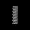 | ||||||||||||||||||||||||
|---|---|---|---|---|---|---|---|---|---|---|---|---|---|---|---|---|---|---|---|---|---|---|---|---|---|
| Title | Cryo-EM structure of the fd bacteriophage capsid major coat protein pVIII | ||||||||||||||||||||||||
 Map data Map data | Cryo-EM map of the intact fd bacteriophage capsid | ||||||||||||||||||||||||
 Sample Sample |
| ||||||||||||||||||||||||
 Keywords Keywords | Bacteriophage / fd / inovirus / ff / helical / phage / filamentous / VIRUS | ||||||||||||||||||||||||
| Function / homology | Phage major coat protein, Gp8 / Bacteriophage M13, G8P, capsid domain superfamily / Capsid protein G8P / helical viral capsid / host cell membrane / membrane / Capsid protein G8P Function and homology information Function and homology information | ||||||||||||||||||||||||
| Biological species |   Enterobacteria phage fd (virus) Enterobacteria phage fd (virus) | ||||||||||||||||||||||||
| Method | helical reconstruction / cryo EM / Resolution: 3.2 Å | ||||||||||||||||||||||||
 Authors Authors | Boehning J / Bharat TAM | ||||||||||||||||||||||||
| Funding support |  United Kingdom, United Kingdom,  France, France,  United States, European Union, 7 items United States, European Union, 7 items
| ||||||||||||||||||||||||
 Citation Citation | Journal: Nat Commun / Year: 2023 Title: Biophysical basis of filamentous phage tactoid-mediated antibiotic tolerance in P. aeruginosa. Authors: Jan Böhning / Miles Graham / Suzanne C Letham / Luke K Davis / Ulrike Schulze / Phillip J Stansfeld / Robin A Corey / Philip Pearce / Abul K Tarafder / Tanmay A M Bharat / Abstract: Inoviruses are filamentous phages infecting numerous prokaryotic phyla. Inoviruses can self-assemble into mesoscale structures with liquid-crystalline order, termed tactoids, which protect bacterial ...Inoviruses are filamentous phages infecting numerous prokaryotic phyla. Inoviruses can self-assemble into mesoscale structures with liquid-crystalline order, termed tactoids, which protect bacterial cells in Pseudomonas aeruginosa biofilms from antibiotics. Here, we investigate the structural, biophysical, and protective properties of tactoids formed by the P. aeruginosa phage Pf4 and Escherichia coli phage fd. A cryo-EM structure of the capsid from fd revealed distinct biochemical properties compared to Pf4. Fd and Pf4 formed tactoids with different morphologies that arise from differing phage geometries and packing densities, which in turn gave rise to different tactoid emergent properties. Finally, we showed that tactoids formed by either phage protect rod-shaped bacteria from antibiotic treatment, and that direct association with a tactoid is required for protection, demonstrating the formation of a diffusion barrier by the tactoid. This study provides insights into how filamentous molecules protect bacteria from extraneous substances in biofilms and in host-associated infections. #1: Journal: Biorxiv / Year: 2023Title: Biophysical basis of phage liquid crystalline droplet-mediated antibiotic tolerance in pathogenic bacteria Authors: Bohning J / Graham M / Letham S / Davis L / Schulze U / Stansfeld P / Corey R / Pearce P / Tarafder A / Bharat T | ||||||||||||||||||||||||
| History |
|
- Structure visualization
| Supplemental images |
|---|

- Downloads & links
-EMDB archive
| Map data | emd_16657.map.gz | 96.3 MB | EMDB map data format | |
|---|---|---|---|---|
| Header (meta data) | emd-16657-v30.xmlemd-16657.xml | 18.8 KB 18.8 KB | Display Display | EMDB header |
| FSC (resolution estimation) | emd_16657_fsc.xml | 10.6 KB | Display | FSC data file |
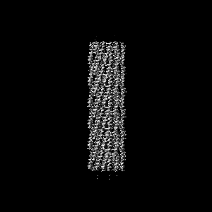
| Images | 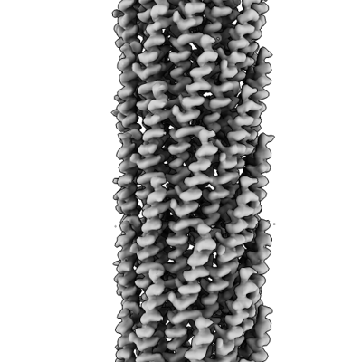 emd_16657.png emd_16657.png | 101.5 KB | ||
| Filedesc metadata | emd-16657.cif.gz | 5.9 KB | ||
| Others | emd_16657_half_map_1.map.gzemd_16657_half_map_2.map.gz | 81.1 MB 80.9 MB | ||
| Archive directory |  http://ftp.pdbj.org/pub/emdb/structures/EMD-16657ftp://ftp.pdbj.org/pub/emdb/structures/EMD-16657 http://ftp.pdbj.org/pub/emdb/structures/EMD-16657ftp://ftp.pdbj.org/pub/emdb/structures/EMD-16657 | HTTPS FTP |
-Validation report
| Summary document | emd_16657_validation.pdf.gz | 997.6 KB | Display | EMDB validaton report |
|---|---|---|---|---|
| Full document | emd_16657_full_validation.pdf.gz | 997.2 KB | Display | |
| Data in XML | emd_16657_validation.xml.gz | 17.8 KB | Display | |
| Data in CIF | emd_16657_validation.cif.gz | 23.4 KB | Display | |
| Arichive directory | https://ftp.pdbj.org/pub/emdb/validation_reports/EMD-16657ftp://ftp.pdbj.org/pub/emdb/validation_reports/EMD-16657 | HTTPS FTP |
-Related structure data
| Related structure data |  8ch5MC M: atomic model generated by this map C: citing same article ( |
|---|---|
| Similar structure data |
-Links
| EMDB pages | EMDB (EBI/PDBe) / EMDataResource |
|---|
-Map
| File | Download / File: emd_16657.map.gz / Format: CCP4 / Size: 103 MB / Type: IMAGE STORED AS FLOATING POINT NUMBER (4 BYTES) | ||||||||||||||||||||||||||||||||||||
|---|---|---|---|---|---|---|---|---|---|---|---|---|---|---|---|---|---|---|---|---|---|---|---|---|---|---|---|---|---|---|---|---|---|---|---|---|---|
| Annotation | Cryo-EM map of the intact fd bacteriophage capsid | ||||||||||||||||||||||||||||||||||||
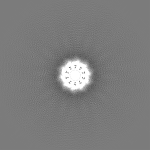
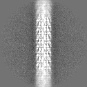
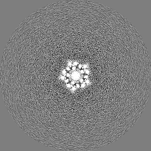
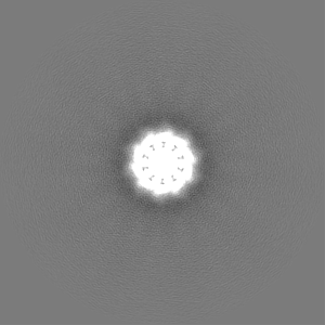
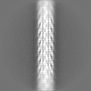
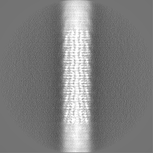
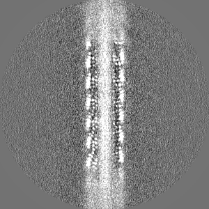
| Projections & slices | Image control
Images are generated by Spider. | ||||||||||||||||||||||||||||||||||||
| Voxel size | X=Y=Z: 1.092 Å | ||||||||||||||||||||||||||||||||||||
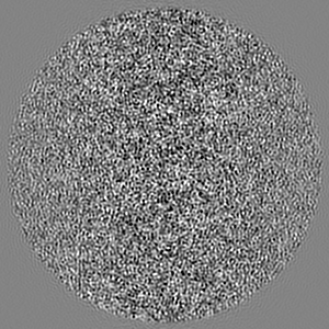
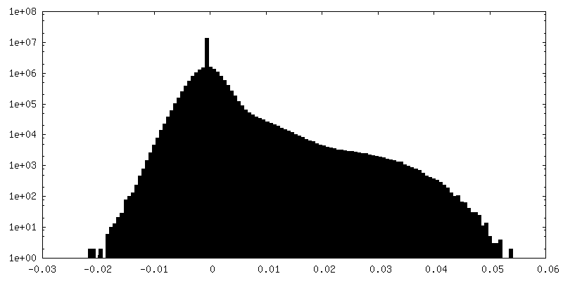
| Density |
| ||||||||||||||||||||||||||||||||||||
| Symmetry | Space group: 1 | ||||||||||||||||||||||||||||||||||||
| Details | EMDB XML:
|
 Z (Sec.)
 Y (Row.)
 X (Col.)


-Supplemental data
-Half map: Half-map 2
| File | emd_16657_half_map_1.map | ||||||||||||
|---|---|---|---|---|---|---|---|---|---|---|---|---|---|
| Annotation | Half-map 2 | ||||||||||||
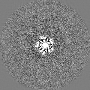
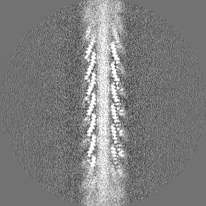
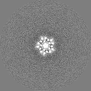
| Projections & Slices |
| ||||||||||||
| Density Histograms |


-Half map: Half-map 1
| File | emd_16657_half_map_2.map | ||||||||||||
|---|---|---|---|---|---|---|---|---|---|---|---|---|---|
| Annotation | Half-map 1 | ||||||||||||
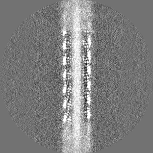
| Projections & Slices |
| ||||||||||||
| Density Histograms |


- Sample components
-Entire : Enterobacteria phage fd
| Entire | Name: Enterobacteria phage fd (virus) |
|---|---|
| Components |
|
-Supramolecule #1: Enterobacteria phage fd
| Supramolecule | Name: Enterobacteria phage fd / type: virus / ID: 1 / Parent: 0 / Macromolecule list: all Details: Purified by PEG precipitation from the supernatant of infected E. coli cells. NCBI-ID: 10864 / Sci species name: Enterobacteria phage fd / Virus type: VIRION / Virus isolate: STRAIN / Virus enveloped: No / Virus empty: No |
|---|---|
| Host (natural) | Organism: |
-Macromolecule #1: Major capsid protein pVIII
| Macromolecule | Name: Major capsid protein pVIII / type: protein_or_peptide / ID: 1 / Number of copies: 1 / Enantiomer: LEVO |
|---|---|
| Source (natural) | Organism: |
| Molecular weight | Theoretical: 5.244 KDa |
| Sequence | String: AEGDDPAKAA FDSLQASATE YIGYAWAMVV VIVGATIGIK LFKKFTSKAS UniProtKB: Capsid protein G8P |
-Experimental details
-Structure determination
| Method | cryo EM |
|---|---|
 Processing Processing | helical reconstruction |
| Aggregation state | filament |
-Sample preparation
| Buffer | pH: 7.4 / Details: Phosphate-buffered saline |
|---|---|
| Grid | Model: Quantifoil R2/2 / Material: COPPER/RHODIUM / Mesh: 200 / Support film - Material: CARBON / Support film - topology: HOLEY ARRAY / Pretreatment - Type: GLOW DISCHARGE / Pretreatment - Time: 20 sec. / Pretreatment - Atmosphere: AIR / Details: 15 mA |
| Vitrification | Cryogen name: ETHANE / Chamber humidity: 100 % / Chamber temperature: 283 K / Instrument: FEI VITROBOT MARK IV |
| Details | fd phage in PBS |
- Electron microscopy
| Microscope | FEI TITAN KRIOS |
|---|---|
| Specialist optics | Energy filter - Name: GIF Bioquantum / Energy filter - Slit width: 20 eV |
| Image recording | Film or detector model: GATAN K3 BIOQUANTUM (6k x 4k) / Detector mode: COUNTING / Number grids imaged: 1 / Average electron dose: 53.9 e/Å2 |
| Electron beam | Acceleration voltage: 300 kV / Electron source:  FIELD EMISSION GUN FIELD EMISSION GUN |
| Electron optics | Illumination mode: FLOOD BEAM / Imaging mode: BRIGHT FIELD / Cs: 2.7 mm / Nominal defocus max: 3.0 µm / Nominal defocus min: 1.0 µm |
| Sample stage | Specimen holder model: FEI TITAN KRIOS AUTOGRID HOLDER / Cooling holder cryogen: NITROGEN |
| Experimental equipment |  Model: Titan Krios / Image courtesy: FEI Company |
-Image processing
| Final reconstruction | Applied symmetry - Helical parameters - Δz: 16.62 Å Applied symmetry - Helical parameters - Δ&Phi: -35.46 ° Applied symmetry - Helical parameters - Axial symmetry: C5 (5 fold cyclic) Resolution.type: BY AUTHOR / Resolution: 3.2 Å / Resolution method: FSC 0.143 CUT-OFF / Software - Name: RELION (ver. 3.1) / Details: Relion 3.1 / Number images used: 84458 |
|---|---|
| Startup model | Type of model: OTHER Details: Cylindrical reference with no internal features (unbiased) |
| Final angle assignment | Type: NOT APPLICABLE |
| FSC plot (resolution estimation) | 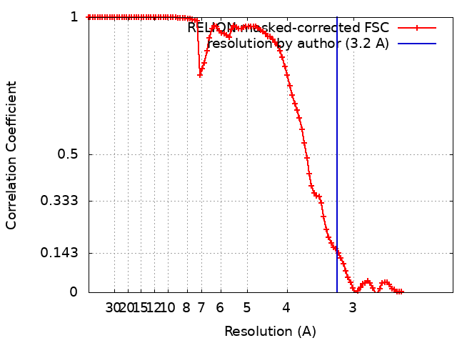 |
-Atomic model buiding 1
| Refinement | Space: REAL / Protocol: AB INITIO MODEL / Overall B value: 79.94 |
|---|---|
| Output model | PDB-8ch5: |
