exoribonuclease II activity / exoribonuclease II / mRNA catabolic process / ribosomal small subunit assembly / ribosomal small subunit biogenesis / small ribosomal subunit / small ribosomal subunit rRNA binding / cytosolic small ribosomal subunit / tRNA binding / rRNA binding ...exoribonuclease II activity / exoribonuclease II / mRNA catabolic process / ribosomal small subunit assembly / ribosomal small subunit biogenesis / small ribosomal subunit / small ribosomal subunit rRNA binding / cytosolic small ribosomal subunit / tRNA binding / rRNA binding / structural constituent of ribosome / ribosome / translation / ribonucleoprotein complex / response to antibiotic / mRNA binding / RNA binding / cytoplasm / cytosol Similarity search - Function
RNase II/RNase R, cold shock domain / Ribonuclease B, N-terminal OB domain / Ribonuclease B OB domain / Cold shock domain / Ribonuclease R / Ribonuclease II/ribonuclease R / Ribonuclease II/R, conserved site / Ribonuclease II family signature. / : / Ribonuclease II/R ...RNase II/RNase R, cold shock domain / Ribonuclease B, N-terminal OB domain / Ribonuclease B OB domain / Cold shock domain / Ribonuclease R / Ribonuclease II/ribonuclease R / Ribonuclease II/R, conserved site / Ribonuclease II family signature. / : / Ribonuclease II/R / RNB domain / RNB / Cold shock domain / Cold shock protein domain / S1 domain profile. / Ribosomal protein S1-like RNA-binding domain / S1 RNA binding domain / Ribosomal protein S16, conserved site / Ribosomal protein S16 signature. / S1 domain / Ribosomal protein S19, bacterial-type / Ribosomal protein S13, bacterial-type / Ribosomal protein S6, conserved site / Ribosomal protein S6 signature. / Ribosomal protein S7, bacterial/organellar-type / Ribosomal protein S11, bacterial-type / Ribosomal protein S20 / Ribosomal protein S20 superfamily / Ribosomal protein S20 / Ribosomal protein S4, bacterial-type / 30S ribosomal protein S17 / Ribosomal protein S5, bacterial-type / Ribosomal protein S6, plastid/chloroplast / Ribosomal protein S9, bacterial/plastid / Ribosomal protein S18, conserved site / Ribosomal protein S18 signature. / Ribosomal protein S16 / Ribosomal protein S16 domain superfamily / Ribosomal protein S16 / Ribosomal protein S15, bacterial-type / Ribosomal protein S6 / Ribosomal protein S6 / Ribosomal protein S6 superfamily / Ribosomal protein S12, bacterial-type / Translation elongation factor EF1B/ribosomal protein S6 / Ribosomal protein S18 / Ribosomal protein S18 / Ribosomal protein S18 superfamily / Ribosomal protein S10, conserved site / Ribosomal protein S10 signature. / Ribosomal protein S15/S19, conserved site / Ribosomal protein S19 signature. / Ribosomal protein S10 / Ribosomal protein S5, N-terminal, conserved site / Ribosomal protein S5 signature. / Ribosomal protein S19/S15 / Ribosomal protein S19/S15, superfamily / Ribosomal protein S19 / Ribosomal protein S7, conserved site / Ribosomal protein S7 signature. / : / Ribosomal protein S5 / S5 double stranded RNA-binding domain profile. / Ribosomal protein S5, N-terminal / Ribosomal protein S5, C-terminal / Ribosomal protein S5, N-terminal domain / Ribosomal protein S17, conserved site / Ribosomal protein S17 signature. / Ribosomal protein S5, C-terminal domain / Ribosomal protein S13, conserved site / Ribosomal protein S13 signature. / Ribosomal protein S13 / 30s ribosomal protein S13, C-terminal / Ribosomal protein S13/S18 / Ribosomal protein S13 family profile. / Ribosomal protein S8 signature. / Ribosomal protein S4/S9 N-terminal domain / Ribosomal protein S4/S9 N-terminal domain / Ribosomal protein S4/S9, N-terminal / Ribosomal protein S4, conserved site / Ribosomal protein S15 signature. / Ribosomal protein S4 signature. / Ribosomal protein S4/S9 / Ribosomal protein S8 / Ribosomal protein S8 superfamily / Ribosomal protein S8 / S4 RNA-binding domain profile. / Ribosomal protein S10p/S20e / Ribosomal protein S13-like, H2TH / Ribosomal protein S10 domain / Ribosomal protein S10 domain superfamily / Ribosomal protein S10p/S20e / Ribosomal S11, conserved site / Ribosomal protein S11 signature. / S4 RNA-binding domain / S4 domain / Ribosomal protein S11 / Ribosomal protein S9, conserved site / Ribosomal protein S9 signature. / RNA-binding S4 domain Similarity search - Domain/homology
Ribonuclease R / Small ribosomal subunit protein uS11 / Small ribosomal subunit protein uS17 / Small ribosomal subunit protein uS8 / Small ribosomal subunit protein uS13 / Small ribosomal subunit protein uS4 / Small ribosomal subunit protein uS5 / Small ribosomal subunit protein bS6 / Small ribosomal subunit protein uS7 / Small ribosomal subunit protein uS9 ...Ribonuclease R / Small ribosomal subunit protein uS11 / Small ribosomal subunit protein uS17 / Small ribosomal subunit protein uS8 / Small ribosomal subunit protein uS13 / Small ribosomal subunit protein uS4 / Small ribosomal subunit protein uS5 / Small ribosomal subunit protein bS6 / Small ribosomal subunit protein uS7 / Small ribosomal subunit protein uS9 / Small ribosomal subunit protein uS10 / Small ribosomal subunit protein uS12 / Small ribosomal subunit protein uS15 / Small ribosomal subunit protein bS16 / Small ribosomal subunit protein bS18 / Small ribosomal subunit protein uS19 / Small ribosomal subunit protein bS20 Similarity search - Component
Biological species
Bacillus subtilis subsp. subtilis str. 168 (bacteria)
Method
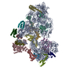
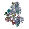
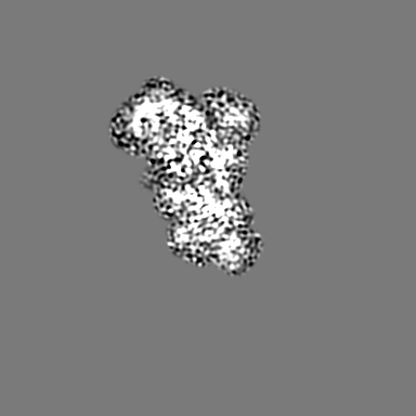
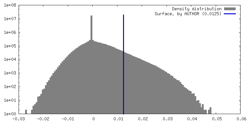
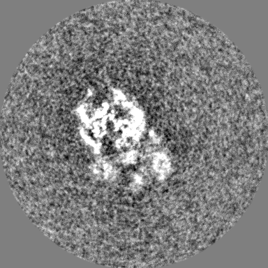
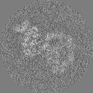
single particle reconstruction / cryo EM / Resolution: 4.73 Å
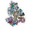
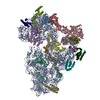
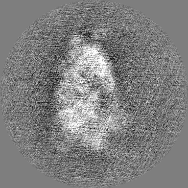
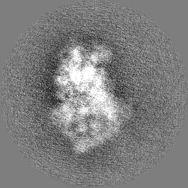
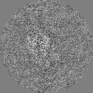
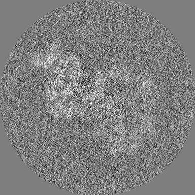
Journal: Nature / Year: 2024 Title: Structural basis of ribosomal 30S subunit degradation by RNase R. Authors: Lyudmila Dimitrova-Paternoga / Sergo Kasvandik / Bertrand Beckert / Sander Granneman / Tanel Tenson / Daniel N Wilson / Helge Paternoga / Abstract: Protein synthesis is a major energy-consuming process of the cell that requires the controlled production and turnover of ribosomes. Although the past few years have seen major advances in our ...Protein synthesis is a major energy-consuming process of the cell that requires the controlled production and turnover of ribosomes. Although the past few years have seen major advances in our understanding of ribosome biogenesis, structural insight into the degradation of ribosomes has been lacking. Here we present native structures of two distinct small ribosomal 30S subunit degradation intermediates associated with the 3' to 5' exonuclease ribonuclease R (RNase R). The structures reveal that RNase R binds at first to the 30S platform to facilitate the degradation of the functionally important anti-Shine-Dalgarno sequence and the decoding-site helix 44. RNase R then encounters a roadblock when it reaches the neck region of the 30S subunit, and this is overcome by a major structural rearrangement of the 30S head, involving the loss of ribosomal proteins. RNase R parallels this movement and relocates to the decoding site by using its N-terminal helix-turn-helix domain as an anchor. In vitro degradation assays suggest that head rearrangement poses a major kinetic barrier for RNase R, but also indicate that the enzyme alone is sufficient for complete degradation of 30S subunits. Collectively, our results provide a mechanistic basis for the degradation of 30S mediated by RNase R, and reveal that RNase R targets orphaned 30S subunits using a dynamic mechanism involving an anchored switching of binding sites.
In the structure databanks used in Yorodumi, some data are registered as the other names, "COVID-19 virus" and "2019-nCoV". Here are the details of the virus and the list of structure data.
Jan 31, 2019. EMDB accession codes are about to change! (news from PDBe EMDB page)
EMDB accession codes are about to change! (news from PDBe EMDB page)
The allocation of 4 digits for EMDB accession codes will soon come to an end. Whilst these codes will remain in use, new EMDB accession codes will include an additional digit and will expand incrementally as the available range of codes is exhausted. The current 4-digit format prefixed with “EMD-” (i.e. EMD-XXXX) will advance to a 5-digit format (i.e. EMD-XXXXX), and so on. It is currently estimated that the 4-digit codes will be depleted around Spring 2019, at which point the 5-digit format will come into force.
The EM Navigator/Yorodumi systems omit the EMD- prefix.
Related info.:Q: What is EMD? / ID/Accession-code notation in Yorodumi/EM Navigator
Yorodumi is a browser for structure data from EMDB, PDB, SASBDB, etc.
This page is also the successor to EM Navigator detail page, and also detail information page/front-end page for Omokage search.
The word "yorodu" (or yorozu) is an old Japanese word meaning "ten thousand". "mi" (miru) is to see.
Related info.:EMDB / PDB / SASBDB / Comparison of 3 databanks / Yorodumi Search / Aug 31, 2016. New EM Navigator & Yorodumi / Yorodumi Papers / Jmol/JSmol / Function and homology information / Changes in new EM Navigator and Yorodumi
 Movie
Movie Controller


 Open data
 Basic information
 Map data
 Sample
 Keywords
 Function and homology information
 Authors
 Germany, 1 items
  Citation


 Structure visualization
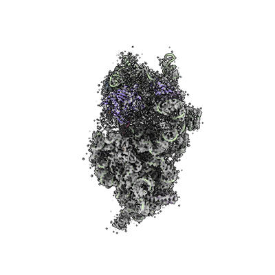
 Downloads & links
 emd_16596.png
 http://ftp.pdbj.org/pub/emdb/structures/EMD-16596
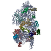


 Z (Sec.)
 Y (Row.)
 X (Col.)


 Sample components
 Processing
 Electron microscopy
 FIELD EMISSION GUN

