Movie
Movie Controller

[English] 日本語
 Yorodumi
- EMDB-16533: Cryo-EM structure of native Otu2-bound ubiquitinated 48S initiati... -
+ Open data

- Basic information
| Entry |  | ||||||||||||||||||
|---|---|---|---|---|---|---|---|---|---|---|---|---|---|---|---|---|---|---|---|
| Title | Cryo-EM structure of native Otu2-bound ubiquitinated 48S initiation complex (partial) | ||||||||||||||||||
 Map data Map data | Cryo-EM map of Otu2-bound ubiquitinated 48S initiation complex | ||||||||||||||||||
 Sample Sample |
| ||||||||||||||||||
 Keywords Keywords | Ribosome / Translation / Ubiquitin / Deubiquitinating enzyme / Initiation / Complex | ||||||||||||||||||
| Function / homology |  Function and homology information Function and homology informationeukaryotic translation initiation factor 3 complex, eIF3e / eukaryotic translation initiation factor 3 complex, eIF3m / translation reinitiation / incipient cellular bud site / multi-eIF complex / formation of cytoplasmic translation initiation complex / eukaryotic translation initiation factor 3 complex / protein-synthesizing GTPase / eukaryotic 43S preinitiation complex / maturation of SSU-rRNA from tricistronic rRNA transcript (SSU-rRNA, LSU-rRNA,5S) ...eukaryotic translation initiation factor 3 complex, eIF3e / eukaryotic translation initiation factor 3 complex, eIF3m / translation reinitiation / incipient cellular bud site / multi-eIF complex / formation of cytoplasmic translation initiation complex / eukaryotic translation initiation factor 3 complex / protein-synthesizing GTPase / eukaryotic 43S preinitiation complex / maturation of SSU-rRNA from tricistronic rRNA transcript (SSU-rRNA, LSU-rRNA,5S) / eukaryotic 48S preinitiation complex / negative regulation of glucose mediated signaling pathway / negative regulation of translational frameshifting / Negative regulators of DDX58/IFIH1 signaling / positive regulation of translational fidelity / Protein methylation / RMTs methylate histone arginines / mTORC1-mediated signalling / Protein hydroxylation / ribosome-associated ubiquitin-dependent protein catabolic process / GDP-dissociation inhibitor activity / nonfunctional rRNA decay / positive regulation of nuclear-transcribed mRNA catabolic process, deadenylation-dependent decay / Formation of the ternary complex, and subsequently, the 43S complex / Translation initiation complex formation / Ribosomal scanning and start codon recognition / preribosome, small subunit precursor / mRNA destabilization / Major pathway of rRNA processing in the nucleolus and cytosol / SRP-dependent cotranslational protein targeting to membrane / GTP hydrolysis and joining of the 60S ribosomal subunit / Formation of a pool of free 40S subunits / Nonsense Mediated Decay (NMD) independent of the Exon Junction Complex (EJC) / Nonsense Mediated Decay (NMD) enhanced by the Exon Junction Complex (EJC) / L13a-mediated translational silencing of Ceruloplasmin expression / regulation of cellular amino acid metabolic process / ribosomal large subunit export from nucleus / G-protein alpha-subunit binding / endonucleolytic cleavage to generate mature 3'-end of SSU-rRNA from (SSU-rRNA, 5.8S rRNA, LSU-rRNA) / positive regulation of protein kinase activity / regulation of translational fidelity / Ub-specific processing proteases / translation regulator activity / ribosomal subunit export from nucleus / ribosomal small subunit export from nucleus / endonucleolytic cleavage in ITS1 to separate SSU-rRNA from 5.8S rRNA and LSU-rRNA from tricistronic rRNA transcript (SSU-rRNA, 5.8S rRNA, LSU-rRNA) / DNA-(apurinic or apyrimidinic site) endonuclease activity / translation initiation factor binding / translation initiation factor activity / 90S preribosome / cellular response to amino acid starvation / rescue of stalled ribosome / maturation of LSU-rRNA from tricistronic rRNA transcript (SSU-rRNA, 5.8S rRNA, LSU-rRNA) / ribosome assembly / maturation of SSU-rRNA from tricistronic rRNA transcript (SSU-rRNA, 5.8S rRNA, LSU-rRNA) / maturation of SSU-rRNA / small-subunit processome / translational initiation / protein kinase C binding / maintenance of translational fidelity / modification-dependent protein catabolic process / cytoplasmic stress granule / rRNA processing / protein tag activity / ribosome biogenesis / ribosome binding / ribosomal small subunit biogenesis / ribosomal small subunit assembly / small ribosomal subunit / small ribosomal subunit rRNA binding / cytosolic small ribosomal subunit / cysteine-type deubiquitinase activity / cytosolic large ribosomal subunit / cytoplasmic translation / tRNA binding / rRNA binding / negative regulation of translation / ribosome / protein ubiquitination / structural constituent of ribosome / translation / positive regulation of protein phosphorylation / G protein-coupled receptor signaling pathway / negative regulation of gene expression / GTPase activity / mRNA binding / ubiquitin protein ligase binding / nucleolus / GTP binding / ATP hydrolysis activity / mitochondrion / RNA binding / zinc ion binding / nucleoplasm / ATP binding / nucleus / metal ion binding / cytosol / cytoplasm Similarity search - Function | ||||||||||||||||||
| Biological species |  | ||||||||||||||||||
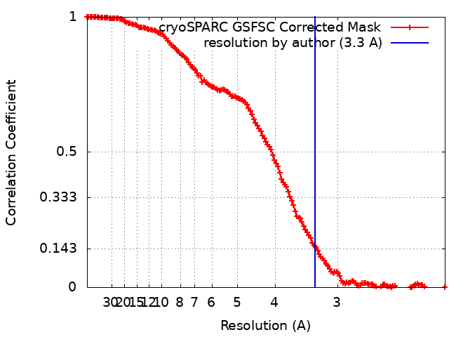
| Method | single particle reconstruction / cryo EM / Resolution: 3.3 Å | ||||||||||||||||||
 Authors Authors | Ikeuchi K / Buschauer R / Cheng J / Berninghausen O / Becker T / Beckmann R | ||||||||||||||||||
| Funding support |  Germany, Germany,  Japan, 5 items Japan, 5 items
| ||||||||||||||||||
 Citation Citation | Journal: Nat Commun / Year: 2023 Title: Molecular basis for recognition and deubiquitination of 40S ribosomes by Otu2. Authors: Ken Ikeuchi / Nives Ivic / Robert Buschauer / Jingdong Cheng / Thomas Fröhlich / Yoshitaka Matsuo / Otto Berninghausen / Toshifumi Inada / Thomas Becker / Roland Beckmann /   Abstract: In actively translating 80S ribosomes the ribosomal protein eS7 of the 40S subunit is monoubiquitinated by the E3 ligase Not4 and deubiquitinated by Otu2 upon ribosomal subunit recycling. Despite its ...In actively translating 80S ribosomes the ribosomal protein eS7 of the 40S subunit is monoubiquitinated by the E3 ligase Not4 and deubiquitinated by Otu2 upon ribosomal subunit recycling. Despite its importance for translation efficiency the exact role and structural basis for this translational reset is poorly understood. Here, structural analysis by cryo-electron microscopy of native and reconstituted Otu2-bound ribosomal complexes reveals that Otu2 engages 40S subunits mainly between ribosome recycling and initiation stages. Otu2 binds to several sites on the intersubunit surface of the 40S that are not occupied by any other 40S-binding factors. This binding mode explains the discrimination against 80S ribosomes via the largely helical N-terminal domain of Otu2 as well as the specificity for mono-ubiquitinated eS7 on 40S. Collectively, this study reveals mechanistic insights into the Otu2-driven deubiquitination steps for translational reset during ribosome recycling/(re)initiation. | ||||||||||||||||||
| History |
|
- Structure visualization
| Supplemental images |
|---|

- Downloads & links
-EMDB archive
| Map data | emd_16533.map.gz | 19.2 MB | EMDB map data format | |
|---|---|---|---|---|
| Header (meta data) | emd-16533-v30.xmlemd-16533.xml | 72.2 KB 72.2 KB | Display Display | EMDB header |
| FSC (resolution estimation) | emd_16533_fsc.xml | 13.9 KB | Display | FSC data file |
| Images |  emd_16533.png emd_16533.png | 160.8 KB | ||
| Filedesc metadata | emd-16533.cif.gz | 16.6 KB | ||
| Others | emd_16533_half_map_1.map.gzemd_16533_half_map_2.map.gz | 261.9 MB 261.9 MB | ||
| Archive directory |  http://ftp.pdbj.org/pub/emdb/structures/EMD-16533ftp://ftp.pdbj.org/pub/emdb/structures/EMD-16533 http://ftp.pdbj.org/pub/emdb/structures/EMD-16533ftp://ftp.pdbj.org/pub/emdb/structures/EMD-16533 | HTTPS FTP |
-Related structure data
| Related structure data |  8casMC  8c83C  8cahC  8cbjC C: citing same article ( M: atomic model generated by this map |
|---|---|
| Similar structure data |


-Links
| EMDB pages | EMDB (EBI/PDBe) / EMDataResource |
|---|---|
| Related items in Molecule of the Month |


-Map
| File | Download / File: emd_16533.map.gz / Format: CCP4 / Size: 282.6 MB / Type: IMAGE STORED AS FLOATING POINT NUMBER (4 BYTES) | ||||||||||||||||||||
|---|---|---|---|---|---|---|---|---|---|---|---|---|---|---|---|---|---|---|---|---|---|
| Annotation | Cryo-EM map of Otu2-bound ubiquitinated 48S initiation complex | ||||||||||||||||||||
| Voxel size | X=Y=Z: 1.045 Å | ||||||||||||||||||||
| Density |
| ||||||||||||||||||||
| Symmetry | Space group: 1 | ||||||||||||||||||||
| Details | EMDB XML:
|
-Supplemental data
-Half map: Half-map A
| File | emd_16533_half_map_1.map | ||||||||||||
|---|---|---|---|---|---|---|---|---|---|---|---|---|---|
| Annotation | Half-map A | ||||||||||||
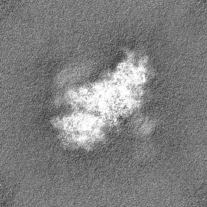
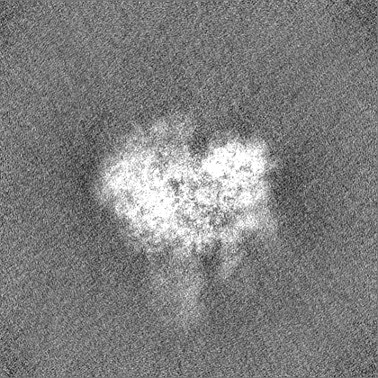
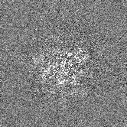
| Projections & Slices |
| ||||||||||||
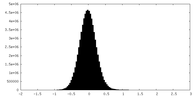
| Density Histograms |
 Z
Z Y
Y X
X


-Half map: Half-map B
| File | emd_16533_half_map_2.map | ||||||||||||
|---|---|---|---|---|---|---|---|---|---|---|---|---|---|
| Annotation | Half-map B | ||||||||||||
| Projections & Slices |
| ||||||||||||
| Density Histograms |


- Sample components
+Entire : Yeast native 48S initiation complex (partial) with deubiquitinati...
+Supramolecule #1: Yeast native 48S initiation complex (partial) with deubiquitinati...
+Macromolecule #1: 40S ribosomal protein S0-A
+Macromolecule #2: 40S ribosomal protein S1-A
+Macromolecule #3: 40S ribosomal protein S2
+Macromolecule #4: 40S ribosomal protein S4-B
+Macromolecule #5: 40S ribosomal protein S6-B
+Macromolecule #6: 40S ribosomal protein S8-A
+Macromolecule #7: 40S ribosomal protein S9-A
+Macromolecule #8: 40S ribosomal protein S11-A
+Macromolecule #9: 40S ribosomal protein S13
+Macromolecule #10: 40S ribosomal protein S14-A
+Macromolecule #11: 40S ribosomal protein S21-A
+Macromolecule #12: 40S ribosomal protein S22-A
+Macromolecule #13: 40S ribosomal protein S23-A
+Macromolecule #14: 40S ribosomal protein S24-A
+Macromolecule #15: 40S ribosomal protein S26-A
+Macromolecule #16: 40S ribosomal protein S27-A
+Macromolecule #17: 40S ribosomal protein S30-A
+Macromolecule #18: RPS15 isoform 1
+Macromolecule #19: 40S ribosomal protein S3
+Macromolecule #20: 40S ribosomal protein S10-A
+Macromolecule #21: 40S ribosomal protein S12
+Macromolecule #22: 40S ribosomal protein S16-A
+Macromolecule #23: 40S ribosomal protein S17-A
+Macromolecule #24: 40S ribosomal protein S18-A
+Macromolecule #25: 40S ribosomal protein S19-A
+Macromolecule #26: 40S ribosomal protein S20
+Macromolecule #27: 40S ribosomal protein S25-A
+Macromolecule #28: RPS29A isoform 1
+Macromolecule #29: 40S ribosomal protein S31
+Macromolecule #30: Guanine nucleotide-binding protein subunit beta-like protein
+Macromolecule #31: 40S ribosomal protein S28-A
+Macromolecule #32: Eukaryotic translation initiation factor 3 subunit I
+Macromolecule #33: Eukaryotic translation initiation factor 3 subunit G
+Macromolecule #35: TIF5 isoform 1
+Macromolecule #36: Eukaryotic translation initiation factor 3 subunit A
+Macromolecule #37: Eukaryotic translation initiation factor 3 subunit B
+Macromolecule #38: Eukaryotic translation initiation factor 3 subunit C
+Macromolecule #40: Eukaryotic translation initiation factor 4C
+Macromolecule #41: RLI1 isoform 1
+Macromolecule #43: 40S ribosomal protein S5
+Macromolecule #44: 60S ribosomal protein L40-A
+Macromolecule #45: 60S ribosomal protein L41-A
+Macromolecule #46: SUI2 isoform 1
+Macromolecule #47: protein-synthesizing GTPase
+Macromolecule #48: SUI3 isoform 1
+Macromolecule #49: OTU domain-containing protein 2
+Macromolecule #50: 40S ribosomal protein S7-A
+Macromolecule #34: tRNA
+Macromolecule #39: mRNA
+Macromolecule #42: 18S ribosomal RNA
+Macromolecule #51: ZINC ION
+Macromolecule #52: ADENOSINE-5'-DIPHOSPHATE

+Macromolecule #53: MAGNESIUM ION
+Macromolecule #54: ADENOSINE-5'-TRIPHOSPHATE

+Macromolecule #55: IRON/SULFUR CLUSTER

+Macromolecule #56: METHIONINE

+Macromolecule #57: PHOSPHOMETHYLPHOSPHONIC ACID GUANYLATE ESTER

-Experimental details
-Structure determination
| Method | cryo EM |
|---|---|
 Processing Processing | single particle reconstruction |
| Aggregation state | particle |
-Sample preparation
| Buffer | pH: 7.5 |
|---|---|
| Vitrification | Cryogen name: ETHANE |
- Electron microscopy
| Microscope | FEI TITAN KRIOS |
|---|---|
| Image recording | Film or detector model: GATAN K2 SUMMIT (4k x 4k) / Detector mode: COUNTING / Average electron dose: 46.4 e/Å2 |
| Electron beam | Acceleration voltage: 300 kV / Electron source:  FIELD EMISSION GUN FIELD EMISSION GUN |
| Electron optics | Illumination mode: FLOOD BEAM / Imaging mode: BRIGHT FIELD / Nominal defocus max: 3.0 µm / Nominal defocus min: 0.5 µm |
| Experimental equipment |  Model: Titan Krios / Image courtesy: FEI Company |