 Movie
Movie Controller

[English] 日本語
 Yorodumi
- EMDB-16349: Enp1TAP_A population of yeast small ribosomal subunit precursors,... -
+ Open data

- Basic information
| Entry |  | |||||||||
|---|---|---|---|---|---|---|---|---|---|---|
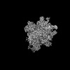
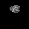
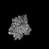
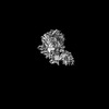
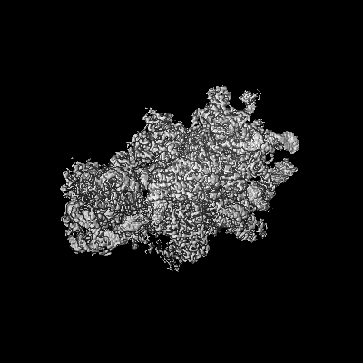
| Title | Enp1TAP_A population of yeast small ribosomal subunit precursors, multibody refinement | |||||||||
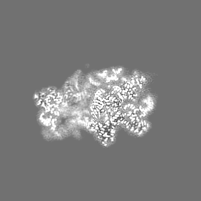
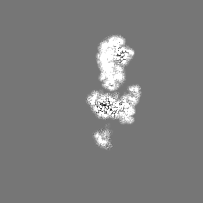
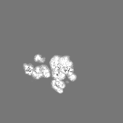
 Map data Map data | Enp1TAP_A Multibody Refinement | |||||||||
 Sample Sample |
| |||||||||
 Keywords Keywords | ribosomal assembly state / RIBOSOME | |||||||||
| Function / homology |  Function and homology information Function and homology information: / positive regulation of RNA import into nucleus / endonucleolytic cleavage of tricistronic rRNA transcript (SSU-rRNA, 5.8S rRNA, LSU-rRNA) / endonucleolytic cleavage to generate mature 5'-end of SSU-rRNA from (SSU-rRNA, 5.8S rRNA, LSU-rRNA) / Negative regulators of DDX58/IFIH1 signaling / rRNA primary transcript binding / Protein methylation / positive regulation of translational fidelity / RMTs methylate histone arginines / mTORC1-mediated signalling ...: / positive regulation of RNA import into nucleus / endonucleolytic cleavage of tricistronic rRNA transcript (SSU-rRNA, 5.8S rRNA, LSU-rRNA) / endonucleolytic cleavage to generate mature 5'-end of SSU-rRNA from (SSU-rRNA, 5.8S rRNA, LSU-rRNA) / Negative regulators of DDX58/IFIH1 signaling / rRNA primary transcript binding / Protein methylation / positive regulation of translational fidelity / RMTs methylate histone arginines / mTORC1-mediated signalling / Protein hydroxylation / U3 snoRNA binding / positive regulation of nuclear-transcribed mRNA catabolic process, deadenylation-dependent decay / Formation of the ternary complex, and subsequently, the 43S complex / poly(A)+ mRNA export from nucleus / Translation initiation complex formation / preribosome, small subunit precursor / Ribosomal scanning and start codon recognition / snoRNA binding / Major pathway of rRNA processing in the nucleolus and cytosol / SRP-dependent cotranslational protein targeting to membrane / GTP hydrolysis and joining of the 60S ribosomal subunit / Nonsense Mediated Decay (NMD) independent of the Exon Junction Complex (EJC) / Nonsense Mediated Decay (NMD) enhanced by the Exon Junction Complex (EJC) / Formation of a pool of free 40S subunits / L13a-mediated translational silencing of Ceruloplasmin expression / proteasome assembly / endonucleolytic cleavage to generate mature 3'-end of SSU-rRNA from (SSU-rRNA, 5.8S rRNA, LSU-rRNA) / 90S preribosome / Ub-specific processing proteases / regulation of translational fidelity / ribosomal subunit export from nucleus / ribonucleoprotein complex binding / ribosomal small subunit export from nucleus / endonucleolytic cleavage in ITS1 to separate SSU-rRNA from 5.8S rRNA and LSU-rRNA from tricistronic rRNA transcript (SSU-rRNA, 5.8S rRNA, LSU-rRNA) / RNA endonuclease activity / ribosome assembly / maturation of SSU-rRNA from tricistronic rRNA transcript (SSU-rRNA, 5.8S rRNA, LSU-rRNA) / maturation of SSU-rRNA / small-subunit processome / translational initiation / maintenance of translational fidelity / cytoplasmic stress granule / rRNA processing / unfolded protein binding / ribosomal small subunit biogenesis / ribosome biogenesis / small ribosomal subunit rRNA binding / ribosomal small subunit assembly / small ribosomal subunit / cytosolic small ribosomal subunit / cytoplasmic translation / Hydrolases; Acting on ester bonds / non-specific serine/threonine protein kinase / rRNA binding / protein kinase activity / ribosome / structural constituent of ribosome / translation / protein serine kinase activity / protein serine/threonine kinase activity / GTPase activity / mRNA binding / GTP binding / nucleolus / endoplasmic reticulum / mitochondrion / RNA binding / nucleoplasm / ATP binding / nucleus / metal ion binding / cytosol / cytoplasm Similarity search - Function | |||||||||
| Biological species |  | |||||||||
| Method | single particle reconstruction / cryo EM / Resolution: 2.7 Å | |||||||||
 Authors Authors | Milkereit P / Poell G | |||||||||
| Funding support |  Germany, 1 items Germany, 1 items
| |||||||||
 Citation Citation | Journal: PLoS One / Year: 2023 Title: Impact of the yeast S0/uS2-cluster ribosomal protein rpS21/eS21 on rRNA folding and the architecture of small ribosomal subunit precursors. Authors: Gisela Pöll / Joachim Griesenbeck / Herbert Tschochner / Philipp Milkereit / Abstract: RpS0/uS2, rpS2/uS5, and rpS21/eS21 form a cluster of ribosomal proteins (S0-cluster) at the head-body junction near the central pseudoknot of eukaryotic small ribosomal subunits (SSU). Previous work ...RpS0/uS2, rpS2/uS5, and rpS21/eS21 form a cluster of ribosomal proteins (S0-cluster) at the head-body junction near the central pseudoknot of eukaryotic small ribosomal subunits (SSU). Previous work in yeast indicated that S0-cluster assembly is required for the stabilisation and maturation of SSU precursors at specific post-nucleolar stages. Here, we analysed the role of S0-cluster formation for rRNA folding. Structures of SSU precursors isolated from yeast S0-cluster expression mutants or control strains were analysed by cryogenic electron microscopy. The obtained resolution was sufficient to detect individual 2'-O-methyl RNA modifications using an unbiased scoring approach. The data show how S0-cluster formation enables the initial recruitment of the pre-rRNA processing factor Nob1 in yeast. Furthermore, they reveal hierarchical effects on the pre-rRNA folding pathway, including the final maturation of the central pseudoknot. Based on these structural insights we discuss how formation of the S0-cluster determines at this early cytoplasmic assembly checkpoint if SSU precursors further mature or are degraded. | |||||||||
| History |
|
- Structure visualization
| Supplemental images |
|---|

- Downloads & links
-EMDB archive
| Map data | emd_16349.map.gz | 14.5 MB | EMDB map data format | |
|---|---|---|---|---|
| Header (meta data) | emd-16349-v30.xmlemd-16349.xml | 47.4 KB 47.4 KB | Display Display | EMDB header |
| Images | 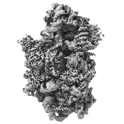 emd_16349.png emd_16349.png | 112.6 KB | ||
| Filedesc metadata | emd-16349.cif.gz | 12 KB | ||
| Archive directory |  http://ftp.pdbj.org/pub/emdb/structures/EMD-16349ftp://ftp.pdbj.org/pub/emdb/structures/EMD-16349 http://ftp.pdbj.org/pub/emdb/structures/EMD-16349ftp://ftp.pdbj.org/pub/emdb/structures/EMD-16349 | HTTPS FTP |
-Validation report
| Summary document | emd_16349_validation.pdf.gz | 405.4 KB | Display | EMDB validaton report |
|---|---|---|---|---|
| Full document | emd_16349_full_validation.pdf.gz | 404.9 KB | Display | |
| Data in XML | emd_16349_validation.xml.gz | 7 KB | Display | |
| Data in CIF | emd_16349_validation.cif.gz | 8.1 KB | Display | |
| Arichive directory | https://ftp.pdbj.org/pub/emdb/validation_reports/EMD-16349ftp://ftp.pdbj.org/pub/emdb/validation_reports/EMD-16349 | HTTPS FTP |
-Related structure data
| Related structure data |  8c01MC  8c00C C: citing same article ( M: atomic model generated by this map |
|---|---|
| Similar structure data |
-Links
| EMDB pages | EMDB (EBI/PDBe) / EMDataResource |
|---|---|
| Related items in Molecule of the Month |


-Map
| File | Download / File: emd_16349.map.gz / Format: CCP4 / Size: 244.1 MB / Type: IMAGE STORED AS FLOATING POINT NUMBER (4 BYTES) | ||||||||||||||||||||||||||||||||||||
|---|---|---|---|---|---|---|---|---|---|---|---|---|---|---|---|---|---|---|---|---|---|---|---|---|---|---|---|---|---|---|---|---|---|---|---|---|---|
| Annotation | Enp1TAP_A Multibody Refinement | ||||||||||||||||||||||||||||||||||||
| Projections & slices | Image control
Images are generated by Spider. | ||||||||||||||||||||||||||||||||||||
| Voxel size | X=Y=Z: 0.968 Å | ||||||||||||||||||||||||||||||||||||
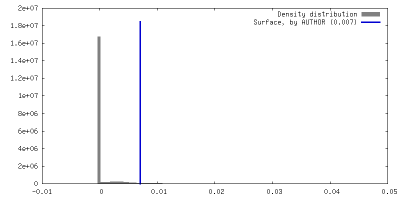
| Density |
| ||||||||||||||||||||||||||||||||||||
| Symmetry | Space group: 1 | ||||||||||||||||||||||||||||||||||||
| Details | EMDB XML:
|
 Z (Sec.)
 Y (Row.)
 X (Col.)


-Supplemental data
- Sample components
+Entire : Enp1-TAP associated immature ribosomal particles from S. cerevisiae
+Supramolecule #1: Enp1-TAP associated immature ribosomal particles from S. cerevisiae
+Macromolecule #1: 18S rRNA precursor
+Macromolecule #2: 40S ribosomal protein S5
+Macromolecule #3: 40S ribosomal protein S17-A
+Macromolecule #4: 40S ribosomal protein S15
+Macromolecule #5: 40S ribosomal protein S16-A
+Macromolecule #6: 40S ribosomal protein S18-A
+Macromolecule #7: 40S ribosomal protein S19-A
+Macromolecule #8: 40S ribosomal protein S25-A
+Macromolecule #9: 40S ribosomal protein S28-A
+Macromolecule #10: 40S ribosomal protein S0-A
+Macromolecule #11: 40S ribosomal protein S1-A
+Macromolecule #12: 40S ribosomal protein S2
+Macromolecule #13: 40S ribosomal protein S4-A
+Macromolecule #14: 40S ribosomal protein S6-A
+Macromolecule #15: 40S ribosomal protein S7-A
+Macromolecule #16: 40S ribosomal protein S8-A
+Macromolecule #17: 40S ribosomal protein S9-A
+Macromolecule #18: 40S ribosomal protein S11-A
+Macromolecule #19: 40S ribosomal protein S13
+Macromolecule #20: 40S ribosomal protein S14-A
+Macromolecule #21: 40S ribosomal protein S21-A
+Macromolecule #22: 40S ribosomal protein S22-A
+Macromolecule #23: 40S ribosomal protein S23-A
+Macromolecule #24: 40S ribosomal protein S24-A
+Macromolecule #25: Essential nuclear protein 1
+Macromolecule #26: 40S ribosomal protein S27-A
+Macromolecule #27: 40S ribosomal protein S30-A
+Macromolecule #28: 20S-pre-rRNA D-site endonuclease NOB1
+Macromolecule #29: Pre-rRNA-processing protein PNO1
+Macromolecule #30: Serine/threonine-protein kinase RIO2
+Macromolecule #31: Ribosome biogenesis protein TSR1
+Macromolecule #32: ZINC ION
-Experimental details
-Structure determination
| Method | cryo EM |
|---|---|
 Processing Processing | single particle reconstruction |
| Aggregation state | particle |
-Sample preparation
| Buffer | pH: 8 Component:
| ||||||||
|---|---|---|---|---|---|---|---|---|---|
| Grid | Model: Quantifoil R1.2/1.3 / Material: COPPER / Mesh: 300 / Support film - Material: CARBON / Support film - topology: HOLEY / Pretreatment - Type: GLOW DISCHARGE / Pretreatment - Time: 200 sec. / Pretreatment - Atmosphere: AIR / Pretreatment - Pressure: 0.4 kPa | ||||||||
| Vitrification | Cryogen name: ETHANE / Chamber humidity: 100 % / Chamber temperature: 277 K / Instrument: FEI VITROBOT MARK IV |
- Electron microscopy
| Microscope | JEOL CRYO ARM 200 |
|---|---|
| Image recording | Film or detector model: GATAN K2 SUMMIT (4k x 4k) / Detector mode: COUNTING / Number grids imaged: 1 / Number real images: 8575 / Average exposure time: 4.4 sec. / Average electron dose: 40.0 e/Å2 |
| Electron beam | Acceleration voltage: 200 kV / Electron source:  FIELD EMISSION GUN FIELD EMISSION GUN |
| Electron optics | C2 aperture diameter: 70.0 µm / Illumination mode: FLOOD BEAM / Imaging mode: BRIGHT FIELD / Cs: 2.7 mm / Nominal defocus max: 2.2 µm / Nominal defocus min: 0.8 µm / Nominal magnification: 50000 |
| Sample stage | Specimen holder model: JEOL CRYOSPECPORTER / Cooling holder cryogen: NITROGEN |
+Image processing
-Atomic model buiding 1
| Refinement | Protocol: FLEXIBLE FIT |
|---|---|
| Output model | PDB-8c01: |
