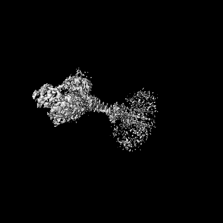
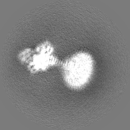
ジャーナル: EMBO Rep / 年: 2024 タイトル: Regulatory sites of CaM-sensitive adenylyl cyclase AC8 revealed by cryo-EM and structural proteomics. 著者: Basavraj Khanppnavar / Dina Schuster / Pia Lavriha / Federico Uliana / Merve Özel / Ved Mehta / Alexander Leitner / Paola Picotti / Volodymyr M Korkhov / 要旨: Membrane adenylyl cyclase AC8 is regulated by G proteins and calmodulin (CaM), mediating the crosstalk between the cAMP pathway and Ca signalling. Despite the importance of AC8 in physiology, the ...Membrane adenylyl cyclase AC8 is regulated by G proteins and calmodulin (CaM), mediating the crosstalk between the cAMP pathway and Ca signalling. Despite the importance of AC8 in physiology, the structural basis of its regulation by G proteins and CaM is not well defined. Here, we report the 3.5 Å resolution cryo-EM structure of the bovine AC8 bound to the stimulatory Gαs protein in the presence of Ca/CaM. The structure reveals the architecture of the ordered AC8 domains bound to Gαs and the small molecule activator forskolin. The extracellular surface of AC8 features a negatively charged pocket, a potential site for unknown interactors. Despite the well-resolved forskolin density, the captured state of AC8 does not favour tight nucleotide binding. The structural proteomics approaches, limited proteolysis and crosslinking mass spectrometry (LiP-MS and XL-MS), allowed us to identify the contact sites between AC8 and its regulators, CaM, Gαs, and Gβγ, as well as to infer the conformational changes induced by these interactions. Our results provide a framework for understanding the role of flexible regions in the mechanism of AC regulation.
全体 : Structure of Adenylyl cyclase 8 bound to stimulatory G protein, F...
全体
名称: Structure of Adenylyl cyclase 8 bound to stimulatory G protein, Forskolin, ATPalphaS, and Ca2+/Calmodulin in lipid nanodisc
要素
複合体: Structure of Adenylyl cyclase 8 bound to stimulatory G protein, Forskolin, ATPalphaS, and Ca2+/Calmodulin in lipid nanodisc
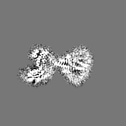
複合体: Adenylyl cyclase 8 and G protein alpha S
タンパク質・ペプチド: Adenylyl cyclase 8
タンパク質・ペプチド: G protein alpha S
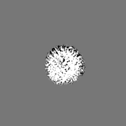
複合体: Calmodulin
タンパク質・ペプチド: Calmodulin
-
超分子 #1: Structure of Adenylyl cyclase 8 bound to stimulatory G protein, F...
超分子
名称: Structure of Adenylyl cyclase 8 bound to stimulatory G protein, Forskolin, ATPalphaS, and Ca2+/Calmodulin in lipid nanodisc タイプ: complex / ID: 1 / 親要素: 0 / 含まれる分子: all
-
超分子 #2: Adenylyl cyclase 8 and G protein alpha S
超分子
名称: Adenylyl cyclase 8 and G protein alpha S / タイプ: complex / ID: 2 / 親要素: 1 / 含まれる分子: #1-#2
 ムービー
ムービー コントローラー


 データを開く
 基本情報
基本情報
 マップデータ
 試料
試料 キーワード
キーワード 機能・相同性情報
 データ登録者
 スイス, 2件
  引用
引用 構造の表示
構造の表示
 ダウンロードとリンク
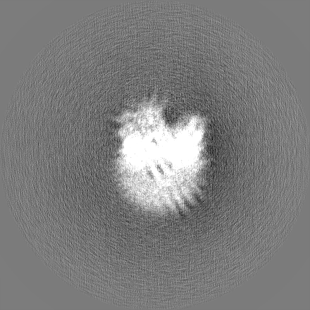
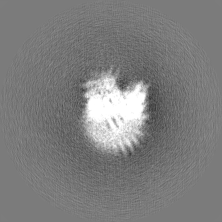
 emd_16249.png
 http://ftp.pdbj.org/pub/emdb/structures/EMD-16249


 Z (Sec.)
 Y (Row.)
 X (Col.)


 試料の構成要素
 Homo sapiens (ヒト)
 Trichoplusia ni (イラクサキンウワバ)
 解析
解析 電子顕微鏡法
 FIELD EMISSION GUN

