 Movie
Movie Controller

[English] 日本語
 Yorodumi
- EMDB-14463: Cryo-EM structure of NNRTI resistant M184I/E138K mutant HIV-1 rev... -
+ Open data

- Basic information
| Entry |  | |||||||||
|---|---|---|---|---|---|---|---|---|---|---|
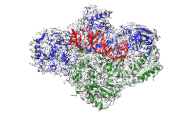
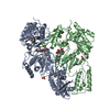
| Title | Cryo-EM structure of NNRTI resistant M184I/E138K mutant HIV-1 reverse transcriptase with a DNA aptamer in complex with rilpivirine | |||||||||
 Map data Map data | ||||||||||
 Sample Sample |
| |||||||||
 Keywords Keywords | Reverse transcriptase mutant / RT-aptamer complex / NNRTI resistance / Non-nucleoside inhibitor / TRANSFERASE | |||||||||
| Function / homology |  Function and homology information Function and homology informationHIV-1 retropepsin / retroviral ribonuclease H / exoribonuclease H / exoribonuclease H activity / host multivesicular body / DNA integration / viral genome integration into host DNA / RNA-directed DNA polymerase / establishment of integrated proviral latency / viral penetration into host nucleus ...HIV-1 retropepsin / retroviral ribonuclease H / exoribonuclease H / exoribonuclease H activity / host multivesicular body / DNA integration / viral genome integration into host DNA / RNA-directed DNA polymerase / establishment of integrated proviral latency / viral penetration into host nucleus / RNA stem-loop binding / RNA-directed DNA polymerase activity / host cell / RNA-DNA hybrid ribonuclease activity / Transferases; Transferring phosphorus-containing groups; Nucleotidyltransferases / symbiont-mediated suppression of host gene expression / viral nucleocapsid / DNA recombination / DNA-directed DNA polymerase / Hydrolases; Acting on ester bonds / aspartic-type endopeptidase activity / DNA-directed DNA polymerase activity / symbiont entry into host cell / lipid binding / host cell nucleus / host cell plasma membrane / structural molecule activity / virion membrane / proteolysis / DNA binding / zinc ion binding / membrane Similarity search - Function | |||||||||
| Biological species |  Human immunodeficiency virus type 1 BH10 / synthetic construct (others) Human immunodeficiency virus type 1 BH10 / synthetic construct (others) | |||||||||
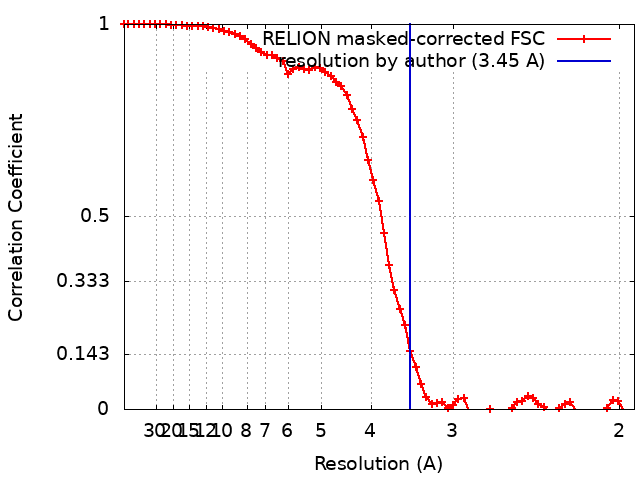
| Method | single particle reconstruction / cryo EM / Resolution: 3.45 Å | |||||||||
 Authors Authors | Singh AK / Das K | |||||||||
| Funding support | 1 items
| |||||||||
 Citation Citation | Journal: Proc Natl Acad Sci U S A / Year: 2022 Title: Cryo-EM structures of wild-type and E138K/M184I mutant HIV-1 RT/DNA complexed with inhibitors doravirine and rilpivirine. Authors: Abhimanyu K Singh / Brent De Wijngaert / Marc Bijnens / Kris Uyttersprot / Hoai Nguyen / Sergio E Martinez / Dominique Schols / Piet Herdewijn / Christophe Pannecouque / Eddy Arnold / Kalyan Das /   Abstract: Structures trapping a variety of functional and conformational states of HIV-1 reverse transcriptase (RT) have been determined by X-ray crystallography. These structures have played important roles ...Structures trapping a variety of functional and conformational states of HIV-1 reverse transcriptase (RT) have been determined by X-ray crystallography. These structures have played important roles in explaining the mechanisms of catalysis, inhibition, and drug resistance and in driving drug design. However, structures of several desired complexes of RT could not be obtained even after many crystallization or crystal soaking experiments. The ternary complexes of doravirine and rilpivirine with RT/DNA are such examples. Structural study of HIV-1 RT by single-particle cryo-electron microscopy (cryo-EM) has been challenging due to the enzyme's relatively smaller size and higher flexibility. We optimized a protocol for rapid structure determination of RT complexes by cryo-EM and determined six structures of wild-type and E138K/M184I mutant RT/DNA in complexes with the nonnucleoside inhibitors rilpivirine, doravirine, and nevirapine. RT/DNA/rilpivirine and RT/DNA/doravirine complexes have structural differences between them and differ from the typical conformation of nonnucleoside RT inhibitor (NNRTI)-bound RT/double-stranded DNA (dsDNA), RT/RNA-DNA, and RT/dsRNA complexes; the primer grip in RT/DNA/doravirine and the YMDD motif in RT/DNA/rilpivirine have large shifts. The DNA primer 3'-end in the doravirine-bound structure is positioned at the active site, but the complex is in a nonproductive state. In the mutant RT/DNA/rilpivirine structure, I184 is stacked with the DNA such that their relative positioning can influence rilpivirine in the pocket. Simultaneously, E138K mutation opens the NNRTI-binding pocket entrance, potentially contributing to a faster rate of rilpivirine dissociation by E138K/M184I mutant RT, as reported by an earlier kinetic study. These structural differences have implications for understanding molecular mechanisms of drug resistance and for drug design. | |||||||||
| History |
|
- Structure visualization
| Supplemental images |
|---|
- Downloads & links
-EMDB archive
| Map data | emd_14463.map.gz | 6.9 MB | EMDB map data format | |
|---|---|---|---|---|
| Header (meta data) | emd-14463-v30.xmlemd-14463.xml | 23.2 KB 23.2 KB | Display Display | EMDB header |
| FSC (resolution estimation) | emd_14463_fsc.xml | 6.9 KB | Display | FSC data file |
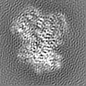
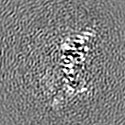
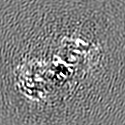
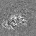
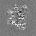
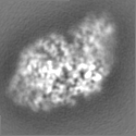
| Images |  emd_14463.png emd_14463.png | 93.5 KB | ||
| Filedesc metadata | emd-14463.cif.gz | 6.9 KB | ||
| Others | emd_14463_half_map_1.map.gzemd_14463_half_map_2.map.gz | 6.9 MB 6.9 MB | ||
| Archive directory |  http://ftp.pdbj.org/pub/emdb/structures/EMD-14463ftp://ftp.pdbj.org/pub/emdb/structures/EMD-14463 http://ftp.pdbj.org/pub/emdb/structures/EMD-14463ftp://ftp.pdbj.org/pub/emdb/structures/EMD-14463 | HTTPS FTP |
-Validation report
| Summary document | emd_14463_validation.pdf.gz | 933.7 KB | Display | EMDB validaton report |
|---|---|---|---|---|
| Full document | emd_14463_full_validation.pdf.gz | 933.2 KB | Display | |
| Data in XML | emd_14463_validation.xml.gz | 12.1 KB | Display | |
| Data in CIF | emd_14463_validation.cif.gz | 15.4 KB | Display | |
| Arichive directory | https://ftp.pdbj.org/pub/emdb/validation_reports/EMD-14463ftp://ftp.pdbj.org/pub/emdb/validation_reports/EMD-14463 | HTTPS FTP |
-Related structure data
| Related structure data | 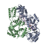 7z2eMC 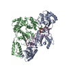 7z24C 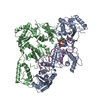 7z29C  7z2dC 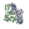 7z2gC 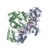 7z2hC C: citing same article ( M: atomic model generated by this map |
|---|---|
| Similar structure data |


-Links
| EMDB pages | EMDB (EBI/PDBe) / EMDataResource |
|---|---|
| Related items in Molecule of the Month |


-Map
| File | Download / File: emd_14463.map.gz / Format: CCP4 / Size: 7.5 MB / Type: IMAGE STORED AS FLOATING POINT NUMBER (4 BYTES) | ||||||||||||||||||||||||||||||||||||
|---|---|---|---|---|---|---|---|---|---|---|---|---|---|---|---|---|---|---|---|---|---|---|---|---|---|---|---|---|---|---|---|---|---|---|---|---|---|
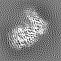
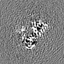
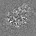
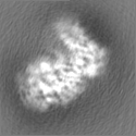
| Projections & slices | Image control
Images are generated by Spider. | ||||||||||||||||||||||||||||||||||||
| Voxel size | X=Y=Z: 0.97 Å | ||||||||||||||||||||||||||||||||||||
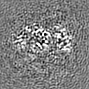
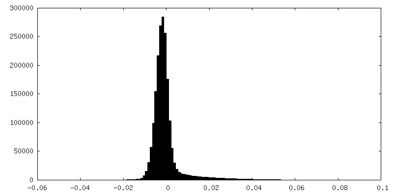
| Density |
| ||||||||||||||||||||||||||||||||||||
| Symmetry | Space group: 1 | ||||||||||||||||||||||||||||||||||||
| Details | EMDB XML:
|
 Z (Sec.)
 Y (Row.)
 X (Col.)


-Supplemental data
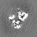
-Half map: #1
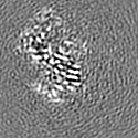
| File | emd_14463_half_map_1.map | ||||||||||||
|---|---|---|---|---|---|---|---|---|---|---|---|---|---|
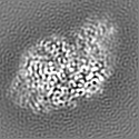
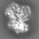
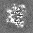
| Projections & Slices |
| ||||||||||||
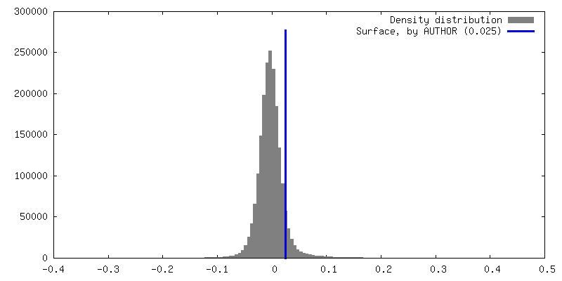
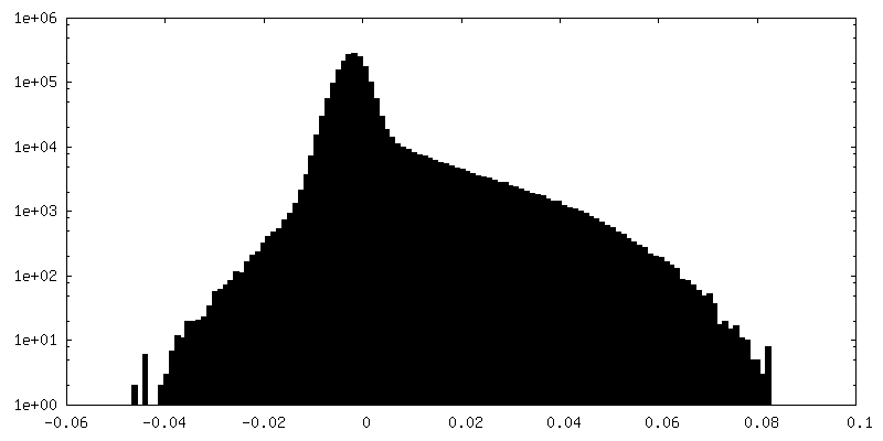
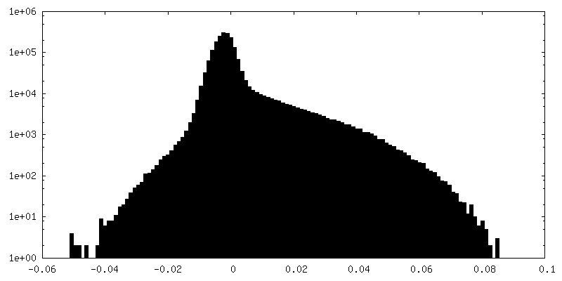
| Density Histograms |


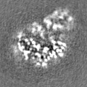
-Half map: #2
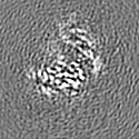
| File | emd_14463_half_map_2.map | ||||||||||||
|---|---|---|---|---|---|---|---|---|---|---|---|---|---|
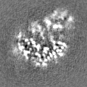
| Projections & Slices |
| ||||||||||||
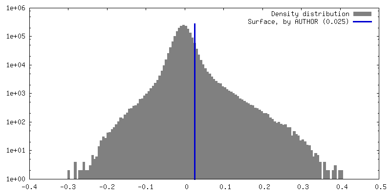
| Density Histograms |


- Sample components
-Entire : NNRTI resistant M184I/E138K mutant HIV-1 reverse transcriptase wi...
| Entire | Name: NNRTI resistant M184I/E138K mutant HIV-1 reverse transcriptase with a DNA aptamer in complex with rilpivirine |
|---|---|
| Components |
|
-Supramolecule #1: NNRTI resistant M184I/E138K mutant HIV-1 reverse transcriptase wi...
| Supramolecule | Name: NNRTI resistant M184I/E138K mutant HIV-1 reverse transcriptase with a DNA aptamer in complex with rilpivirine type: complex / ID: 1 / Parent: 0 / Macromolecule list: #1-#3 |
|---|---|
| Source (natural) | Organism: Human immunodeficiency virus type 1 BH10 |
-Supramolecule #2: HIV-1 REVERSE TRANSCRIPTASE P66 SUBUNIT with M184I mutation
| Supramolecule | Name: HIV-1 REVERSE TRANSCRIPTASE P66 SUBUNIT with M184I mutation type: complex / ID: 2 / Parent: 1 / Macromolecule list: #1 |
|---|---|
| Source (natural) | Organism: Human immunodeficiency virus type 1 BH10 |
-Supramolecule #3: HIV-1 REVERSE TRANSCRIPTASE P51 SUBUNIT with E138K mutation
| Supramolecule | Name: HIV-1 REVERSE TRANSCRIPTASE P51 SUBUNIT with E138K mutation type: complex / ID: 3 / Parent: 1 / Macromolecule list: #2 |
|---|---|
| Source (natural) | Organism: Human immunodeficiency virus type 1 BH10 |
-Macromolecule #1: Reverse transcriptase/ribonuclease H
| Macromolecule | Name: Reverse transcriptase/ribonuclease H / type: protein_or_peptide / ID: 1 / Details: P66 subunit / Number of copies: 1 / Enantiomer: LEVO / EC number: RNA-directed DNA polymerase |
|---|---|
| Source (natural) | Organism: Human immunodeficiency virus type 1 BH10 / Strain: isolate BH10 |
| Molecular weight | Theoretical: 64.019352 KDa |
| Recombinant expression | Organism:  |
| Sequence | String: MVPISPIETV PVKLKPGMDG PKVKQWPLTE EKIKALVEIC TEMEKEGKIS KIGPENPYNT PVFACKKKDS TKWRKLVDFR ELNKRTQDF WEVQLGIPHP AGLKKKKSVT VLDVGDAYFS VPLDEDFRKY TAFTIPSINN ETPGIRYQYN VLPQGWKGSP A IFQSSMTK ...String: MVPISPIETV PVKLKPGMDG PKVKQWPLTE EKIKALVEIC TEMEKEGKIS KIGPENPYNT PVFACKKKDS TKWRKLVDFR ELNKRTQDF WEVQLGIPHP AGLKKKKSVT VLDVGDAYFS VPLDEDFRKY TAFTIPSINN ETPGIRYQYN VLPQGWKGSP A IFQSSMTK ILEPFKKQNP DIVIYQYIDD LYVGSDLEIG QHRTKIEELR QHLLRWGLTT PDKKHQKEPP FLWMGYELHP DK WTVQPIV LPEKDSWTVN DIQKLVGKLN WASQIYPGIK VRQLSKLLRG TKALTEVIPL TEEAELELAE NREILKEPVH GVY YDPSKD LIAEIQKQGQ GQWTYQIYQE PFKNLKTGKY ARMRGAHTND VKQLTEAVQK ITTESIVIWG KTPKFKLPIQ KETW ETWWT EYWQATWIPE WEFVNTPPLV KLWYQLEKEP IVGAETFYVD GAANRETKLG KAGYVTNKGR QKVVPLTNTT NQKTE LQAI YLALQDSGLE VNIVTNSQYA LGIIQAQPDK SESELVNQII EQLIKKEKVY LAWVPAHKGI GGNEQVDKLV SA UniProtKB: Gag-Pol polyprotein |
-Macromolecule #2: p51 RT
| Macromolecule | Name: p51 RT / type: protein_or_peptide / ID: 2 / Details: P51 subunit / Number of copies: 1 / Enantiomer: LEVO |
|---|---|
| Source (natural) | Organism: Human immunodeficiency virus type 1 BH10 / Strain: isolate BH10 |
| Molecular weight | Theoretical: 50.039555 KDa |
| Recombinant expression | Organism: |
| Sequence | String: PISPIETVPV KLKPGMDGPK VKQWPLTEEK IKALVEICTE MEKEGKISKI GPENPYNTPV FAIKKKDSTK WRKLVDFREL NKRTQDFWE VQLGIPHPAG LKKKKSVTVL DVGDAYFSVP LDEDFRKYTA FTIPSINNKT PGIRYQYNVL PQGWKGSPAI F QSSMTKIL ...String: PISPIETVPV KLKPGMDGPK VKQWPLTEEK IKALVEICTE MEKEGKISKI GPENPYNTPV FAIKKKDSTK WRKLVDFREL NKRTQDFWE VQLGIPHPAG LKKKKSVTVL DVGDAYFSVP LDEDFRKYTA FTIPSINNKT PGIRYQYNVL PQGWKGSPAI F QSSMTKIL EPFKKQNPDI VIYQYMDDLY VGSDLEIGQH RTKIEELRQH LLRWGLTTPD KKHQKEPPFL WMGYELHPDK WT VQPIVLP EKDSWTVNDI QKLVGKLNWA SQIYPGIKVR QLSKLLRGTK ALTEVIPLTE EAELELAENR EILKEPVHGV YYD PSKDLI AEIQKQGQGQ WTYQIYQEPF KNLKTGKYAR MRGAHTNDVK QLTEAVQKIT TESIVIWGKT PKFKLPIQKE TWET WWTEY WQATWIPEWE FVNTPPLVKL WYQ UniProtKB: Gag-Pol polyprotein |
-Macromolecule #3: DNA (38-MER)
| Macromolecule | Name: DNA (38-MER) / type: dna / ID: 3 / Number of copies: 1 / Classification: DNA |
|---|---|
| Source (natural) | Organism: synthetic construct (others) |
| Molecular weight | Theoretical: 11.748526 KDa |
| Sequence | String: (DT)(DA)(DA)(DT)(DA)(DC)(OMC)(DC)(OMC)(DC) (DC)(DC)(DT)(DT)(DC)(DG)(DG)(DT)(DG) (DC)(DT)(DT)(DT)(DG)(DC)(DA)(DC)(DC)(DG) (DA)(DA)(DG)(DG)(DG)(DG)(DG)(DG)(DG) |
-Macromolecule #4: 4-{[4-({4-[(E)-2-cyanoethenyl]-2,6-dimethylphenyl}amino)pyrimidin...
| Macromolecule | Name: 4-{[4-({4-[(E)-2-cyanoethenyl]-2,6-dimethylphenyl}amino)pyrimidin-2-yl]amino}benzonitrile type: ligand / ID: 4 / Number of copies: 1 / Formula: T27 |
|---|---|
| Molecular weight | Theoretical: 366.419 Da |
| Chemical component information | 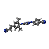 ChemComp-T27: |
-Experimental details
-Structure determination
| Method | cryo EM |
|---|---|
 Processing Processing | single particle reconstruction |
| Aggregation state | particle |
-Sample preparation
| Concentration | 0.3 mg/mL | |||||||||
|---|---|---|---|---|---|---|---|---|---|---|
| Buffer | pH: 8 Component:
| |||||||||
| Grid | Model: Quantifoil R1.2/1.3 / Material: GOLD / Mesh: 200 / Pretreatment - Type: GLOW DISCHARGE / Pretreatment - Time: 60 sec. / Pretreatment - Atmosphere: AIR / Pretreatment - Pressure: 0.03 kPa | |||||||||
| Vitrification | Cryogen name: ETHANE / Chamber humidity: 95 % / Chamber temperature: 281 K / Instrument: LEICA EM GP |
- Electron microscopy
| Microscope | TFS GLACIOS |
|---|---|
| Image recording | Film or detector model: FEI FALCON III (4k x 4k) / Detector mode: COUNTING / Number grids imaged: 1 / Number real images: 1850 / Average exposure time: 55.0 sec. / Average electron dose: 40.0 e/Å2 |
| Electron beam | Acceleration voltage: 200 kV / Electron source:  FIELD EMISSION GUN FIELD EMISSION GUN |
| Electron optics | C2 aperture diameter: 50.0 µm / Calibrated defocus max: 2.5 µm / Calibrated defocus min: 0.5 µm / Illumination mode: FLOOD BEAM / Imaging mode: BRIGHT FIELD / Cs: 2.7 mm / Nominal defocus max: 2.2 µm / Nominal defocus min: 0.8 µm / Nominal magnification: 150000 |
| Sample stage | Cooling holder cryogen: NITROGEN |
+Image processing
-Atomic model buiding 1
| Initial model | PDB ID: Chain - Source name: PDB / Chain - Initial model type: experimental model |
|---|---|
| Refinement | Space: REAL / Protocol: FLEXIBLE FIT / Overall B value: 166.4 / Target criteria: Correlation coefficient |
| Output model | PDB-7z2e: |
