- EMDB-14332: 1.58 A STRUCTURE OF HUMAN APOFERRITIN OBTAINED FROM TITAN KRIOS 2... -
+
データを開く
IDまたはキーワード:
読み込み中...
-
基本情報
登録情報
データベース: EMDB / ID: EMD-14332
タイトル
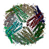
1.58 A STRUCTURE OF HUMAN APOFERRITIN OBTAINED FROM TITAN KRIOS 2 AT eBIC, DLS UNDER COMMISSIONING SESSION CM26464-2
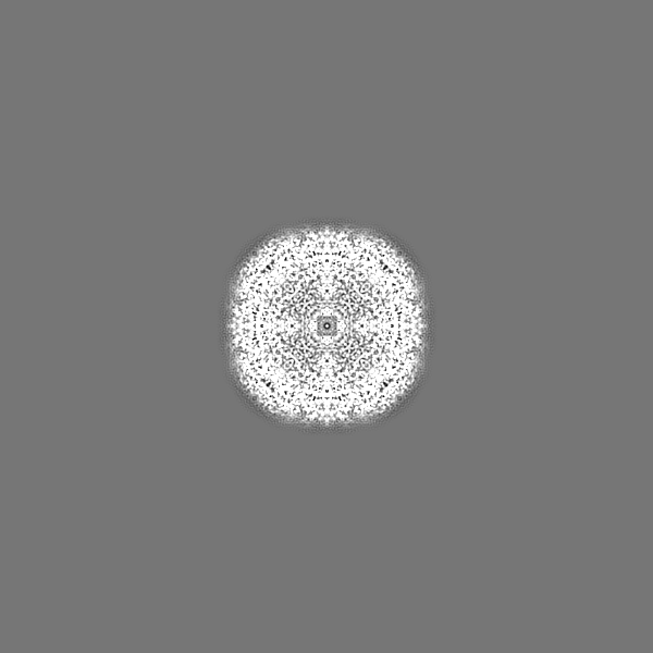
マップデータ
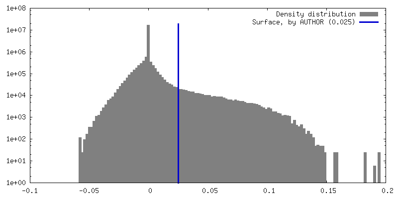
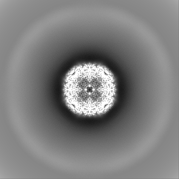
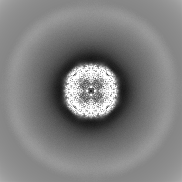
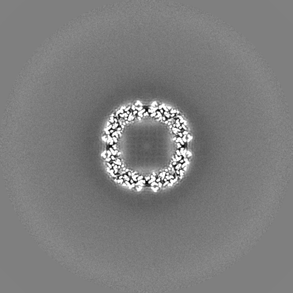
ApoF structure post processed in Relion
試料
複合体: human Apoferritin
タンパク質・ペプチド: Ferritin heavy chain, N-terminally processed
リガンド: SODIUM ION
リガンド: water
キーワード
Protein standard / METAL BINDING PROTEIN
機能・相同性
機能・相同性情報
iron ion sequestering activity / ferritin complex / negative regulation of ferroptosis / Scavenging by Class A Receptors / Golgi Associated Vesicle Biogenesis / ferroxidase / autolysosome / ferroxidase activity / negative regulation of fibroblast proliferation / ferric iron binding ...iron ion sequestering activity / ferritin complex / negative regulation of ferroptosis / Scavenging by Class A Receptors / Golgi Associated Vesicle Biogenesis / ferroxidase / autolysosome / ferroxidase activity / negative regulation of fibroblast proliferation / ferric iron binding / autophagosome / Iron uptake and transport / ferrous iron binding / tertiary granule lumen / iron ion transport / intracellular iron ion homeostasis / ficolin-1-rich granule lumen / immune response / iron ion binding / negative regulation of cell population proliferation / Neutrophil degranulation / extracellular exosome / extracellular region / identical protein binding / nucleus / membrane / cytosol / cytoplasm 類似検索 - 分子機能
ジャーナル: Faraday Discuss / 年: 2022 タイトル: Application of super-resolution and correlative double sampling in cryo-electron microscopy. 著者: Yuewen Sheng / Peter J Harrison / Vinod Vogirala / Zhengyi Yang / Claire Strain-Damerell / Thomas Frosio / Benjamin A Himes / C Alistair Siebert / Peijun Zhang / Daniel K Clare / 要旨: Developments in cryo-EM have allowed atomic or near-atomic resolution structure determination to become routine in single particle analysis (SPA). However, near-atomic resolution structures ...Developments in cryo-EM have allowed atomic or near-atomic resolution structure determination to become routine in single particle analysis (SPA). However, near-atomic resolution structures determined using cryo-electron tomography and sub-tomogram averaging (cryo-ET STA) are much less routine. In this paper, we show that collecting cryo-ET STA data using the same conditions as SPA, with both correlated double sampling (CDS) and the super-resolution mode, allowed apoferritin to be reconstructed out to the physical Nyquist frequency of the images. Even with just two tilt series, STA yields an apoferritin map at 2.9 Å resolution. These results highlight the exciting potential of cryo-ET STA in the future of protein structure determination. While processing SPA data recorded in super-resolution mode may yield structures surpassing the physical Nyquist limit, processing cryo-ET STA data in the super-resolution mode gave no additional resolution benefit. We further show that collecting SPA data in the super-resolution mode, with CDS activated, reduces the estimated -factor, leading to a reduction in the number of particles required to reach a target resolution without compromising the data size on disk and the area imaged in SerialEM. However, collecting SPA data in CDS does reduce throughput, given that a similar resolution structure, with a slightly larger -factor, is achievable with optimised parameters for speed in EPU (without CDS).
 ムービー
ムービー コントローラー


 データを開く
 基本情報
基本情報
 マップデータ
 試料
試料 キーワード
キーワード 機能・相同性情報
 Homo sapiens (ヒト)
 データ登録者
 英国, 1件
  引用
引用
 構造の表示
構造の表示
 ダウンロードとリンク
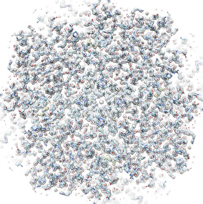 emd_14332.png
 http://ftp.pdbj.org/pub/emdb/structures/EMD-14332


 Z (Sec.)
 Y (Row.)
 X (Col.)


 試料の構成要素

 解析
解析 電子顕微鏡法
 FIELD EMISSION GUN