 Movie
Movie Controller

[English] 日本語
 Yorodumi
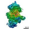
- EMDB-1325: Composition and three-dimensional EM structure of double affinity... -
+ Open data

- Basic information
| Entry | Database: EMDB / ID: EMD-1325 | |||||||||
|---|---|---|---|---|---|---|---|---|---|---|
| Title | Composition and three-dimensional EM structure of double affinity-purified, human prespliceosomal A complexes. | |||||||||
 Map data Map data | Human spliceosomal A complex | |||||||||
 Sample Sample |
| |||||||||
| Biological species |  Homo sapiens (human) Homo sapiens (human) | |||||||||
| Method | single particle reconstruction / cryo EM / Resolution: 45.0 Å | |||||||||
 Authors Authors | Behzadnia N / Golas MM / Hartmuth K / Sander B / Kastner B / Deckert J / Dube P / Will CL / Urlaub H / Stark H / Luhrmann R | |||||||||
 Citation Citation | Journal: EMBO J / Year: 2007 Title: Composition and three-dimensional EM structure of double affinity-purified, human prespliceosomal A complexes. Authors: Nastaran Behzadnia / Monika M Golas / Klaus Hartmuth / Bjoern Sander / Berthold Kastner / Jochen Deckert / Prakash Dube / Cindy L Will / Henning Urlaub / Holger Stark / Reinhard Lührmann /  Abstract: Little is known about the higher-order structure of prespliceosomal A complexes, in which pairing of the pre-mRNA's splice sites occurs. Here, human A complexes were isolated under physiological ...Little is known about the higher-order structure of prespliceosomal A complexes, in which pairing of the pre-mRNA's splice sites occurs. Here, human A complexes were isolated under physiological conditions by double-affinity selection. Purified complexes contained stoichiometric amounts of U1, U2 and pre-mRNA, and crosslinking studies indicated that these form concomitant base pairing interactions with one another. A complexes contained nearly all U1 and U2 proteins plus approximately 50 non-snRNP proteins. Unexpectedly, proteins of the hPrp19/CDC5 complex were also detected, even when A complexes were formed in the absence of U4/U6 snRNPs, demonstrating that they associate independent of the tri-snRNP. Double-affinity purification yielded structurally homogeneous A complexes as evidenced by electron microscopy, and allowed for the first time the generation of a three-dimensional structure. A complexes possess an asymmetric shape (approximately 260 x 200 x 195 angstroms) and contain a main body with various protruding elements, including a head-like domain and foot-like protrusions. Complexes isolated here are well suited for in vitro assembly studies to determine factor requirements for the A to B complex transition. | |||||||||
| History |
|
- Structure visualization
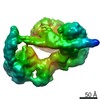
| Movie |
 Movie viewer Movie viewer |
|---|---|
| Structure viewer | EM map: SurfViewMolmilJmol/JSmol |
| Supplemental images |

- Downloads & links
-EMDB archive
| Map data | emd_1325.map.gz | 2 MB | EMDB map data format | |
|---|---|---|---|---|
| Header (meta data) | emd-1325-v30.xmlemd-1325.xml | 7.3 KB 7.3 KB | Display Display | EMDB header |
| Images | 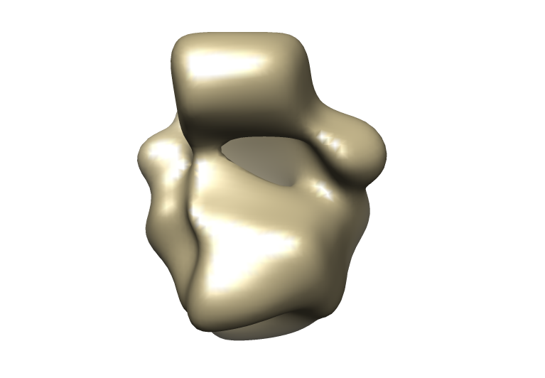 1325.png 1325.png | 99.9 KB | ||
| Archive directory |  http://ftp.pdbj.org/pub/emdb/structures/EMD-1325ftp://ftp.pdbj.org/pub/emdb/structures/EMD-1325 http://ftp.pdbj.org/pub/emdb/structures/EMD-1325ftp://ftp.pdbj.org/pub/emdb/structures/EMD-1325 | HTTPS FTP |
-Validation report
| Summary document | emd_1325_validation.pdf.gz | 184.9 KB | Display | EMDB validaton report |
|---|---|---|---|---|
| Full document | emd_1325_full_validation.pdf.gz | 184 KB | Display | |
| Data in XML | emd_1325_validation.xml.gz | 5 KB | Display | |
| Arichive directory | https://ftp.pdbj.org/pub/emdb/validation_reports/EMD-1325ftp://ftp.pdbj.org/pub/emdb/validation_reports/EMD-1325 | HTTPS FTP |
-Related structure data
| Similar structure data |
|---|


-Links
| EMDB pages | EMDB (EBI/PDBe) / EMDataResource |
|---|
-Map
| File | Download / File: emd_1325.map.gz / Format: CCP4 / Size: 3.7 MB / Type: IMAGE STORED AS FLOATING POINT NUMBER (4 BYTES) | ||||||||||||||||||||||||||||||||||||||||||||||||||||||||||||||||||||
|---|---|---|---|---|---|---|---|---|---|---|---|---|---|---|---|---|---|---|---|---|---|---|---|---|---|---|---|---|---|---|---|---|---|---|---|---|---|---|---|---|---|---|---|---|---|---|---|---|---|---|---|---|---|---|---|---|---|---|---|---|---|---|---|---|---|---|---|---|---|
| Annotation | Human spliceosomal A complex | ||||||||||||||||||||||||||||||||||||||||||||||||||||||||||||||||||||
| Voxel size | X=Y=Z: 4.9 Å | ||||||||||||||||||||||||||||||||||||||||||||||||||||||||||||||||||||
| Density |
| ||||||||||||||||||||||||||||||||||||||||||||||||||||||||||||||||||||
| Symmetry | Space group: 1 | ||||||||||||||||||||||||||||||||||||||||||||||||||||||||||||||||||||
| Details | EMDB XML:
CCP4 map header:
| ||||||||||||||||||||||||||||||||||||||||||||||||||||||||||||||||||||
-Supplemental data
- Sample components
-Entire : Human prespliceosomal A Complex
| Entire | Name: Human prespliceosomal A Complex |
|---|---|
| Components |
|
-Supramolecule #1000: Human prespliceosomal A Complex
| Supramolecule | Name: Human prespliceosomal A Complex / type: sample / ID: 1000 / Number unique components: 1 |
|---|
-Supramolecule #1: Human prespliceosomal A Complex
| Supramolecule | Name: Human prespliceosomal A Complex / type: organelle_or_cellular_component / ID: 1 / Name.synonym: Prespliceosome / Oligomeric state: monomer / Recombinant expression: No |
|---|---|
| Source (natural) | Organism: Homo sapiens (human) / synonym: Human / Cell: HeLa / Organelle: nucleus |
-Experimental details
-Structure determination
| Method | cryo EM |
|---|---|
 Processing Processing | single particle reconstruction |
| Aggregation state | particle |
-Sample preparation
| Vitrification | Cryogen name: ETHANE |
|---|
- Electron microscopy
| Microscope | FEI/PHILIPS CM200FEG |
|---|---|
| Image recording | Category: CCD / Film or detector model: TVIPS TEMCAM-F415 (4k x 4k) |
| Tilt angle min | 0 |
| Electron beam | Acceleration voltage: 200 kV / Electron source:  FIELD EMISSION GUN FIELD EMISSION GUN |
| Electron optics | Calibrated magnification: 122000 / Illumination mode: SPOT SCAN / Imaging mode: BRIGHT FIELD / Nominal magnification: 88000 |
| Sample stage | Specimen holder model: OTHER / Tilt angle max: 45 |
-Image processing
| Final reconstruction | Applied symmetry - Point group: C1 (asymmetric) / Algorithm: OTHER / Resolution.type: BY AUTHOR / Resolution: 45.0 Å / Resolution method: OTHER / Software - Name: IMAGIC |
|---|
