rRNA adenine dimethylase-like, C-terminal / Ribosomal RNA adenine dimethylase / Ribosomal RNA adenine methylase transferase, conserved site / Ribosomal RNA adenine dimethylases signature. / Ribosomal RNA adenine methylase transferase, N-terminal / Ribosomal RNA adenine dimethylases / Ribosomal RNA adenine methyltransferase KsgA/Erm / Ribosomal RNA adenine dimethylase / rRNA adenine N(6)-methyltransferase family profile. / Ribosomal protein S21, conserved site ...rRNA adenine dimethylase-like, C-terminal / Ribosomal RNA adenine dimethylase / Ribosomal RNA adenine methylase transferase, conserved site / Ribosomal RNA adenine dimethylases signature. / Ribosomal RNA adenine methylase transferase, N-terminal / Ribosomal RNA adenine dimethylases / Ribosomal RNA adenine methyltransferase KsgA/Erm / Ribosomal RNA adenine dimethylase / rRNA adenine N(6)-methyltransferase family profile. / Ribosomal protein S21, conserved site / Ribosomal protein S21 signature. / Ribosomal protein S21 superfamily / Ribosomal protein S16, conserved site / Ribosomal protein S16 signature. / Ribosomal protein S21 / Ribosomal protein S21 / Ribosomal protein S6, conserved site / Ribosomal protein S6 signature. / Ribosomal protein S11, bacterial-type / Ribosomal protein S20 / Ribosomal protein S20 superfamily / Ribosomal protein S20 / Ribosomal protein S4, bacterial-type / Ribosomal protein S5, bacterial-type / 30S ribosomal protein S17 / Ribosomal protein S6, plastid/chloroplast / Ribosomal protein S2, bacteria/mitochondria/plastid / Ribosomal protein S18, conserved site / Ribosomal protein S18 signature. / Ribosomal protein S16 / Ribosomal protein S16 domain superfamily / Ribosomal protein S16 / Ribosomal protein S15, bacterial-type / Ribosomal protein S6 / Ribosomal protein S6 / Ribosomal protein S6 superfamily / Ribosomal protein S12, bacterial-type / Translation elongation factor EF1B/ribosomal protein S6 / Ribosomal protein S18 / Ribosomal protein S18 / Ribosomal protein S18 superfamily / Ribosomal protein S2 signature 2. / Ribosomal protein S2 signature 1. / Ribosomal protein S5, N-terminal, conserved site / Ribosomal protein S5 signature. / Ribosomal protein S2, conserved site / Ribosomal protein S2 / Ribosomal protein S2, flavodoxin-like domain superfamily / Ribosomal protein S2 / Ribosomal protein S17, conserved site / Ribosomal protein S17 signature. / Ribosomal protein S5 / S5 double stranded RNA-binding domain profile. / Ribosomal protein S5, N-terminal / Ribosomal protein S5, C-terminal / Ribosomal protein S5, N-terminal domain / Ribosomal protein S5, C-terminal domain / Ribosomal protein S4/S9 N-terminal domain / Ribosomal protein S8 signature. / Ribosomal protein S4, conserved site / Ribosomal protein S4 signature. / Ribosomal protein S4/S9 N-terminal domain / Ribosomal protein S4/S9, N-terminal / Ribosomal protein S15 signature. / Ribosomal protein S4/S9 / Ribosomal protein S8 / Ribosomal protein S8 superfamily / Ribosomal protein S8 / S4 RNA-binding domain profile. / S4 RNA-binding domain / Ribosomal S11, conserved site / S4 domain / Ribosomal protein S11 signature. / RNA-binding S4 domain / RNA-binding S4 domain superfamily / Ribosomal protein S11 / Ribosomal protein S12 signature. / Ribosomal protein S11 / Ribosomal protein S12/S23 / Ribosomal protein S12/S23 / Ribosomal protein S17/S11 / Ribosomal protein S17 / Ribosomal protein S15 / Ribosomal_S15 / Ribosomal protein S15 / Ribosomal protein S11 superfamily / S15/NS1, RNA-binding / Ribosomal protein S5 domain 2-type fold, subgroup / Ribosomal protein S5 domain 2-type fold / S-adenosyl-L-methionine-dependent methyltransferase superfamily / Nucleic acid-binding, OB-fold 類似検索 - ドメイン・相同性
30S ribosomal protein S2 / Ribosomal RNA small subunit methyltransferase A / 30S ribosomal protein S21 / Small ribosomal subunit protein uS11 / Small ribosomal subunit protein uS5 / Small ribosomal subunit protein bS18 / Small ribosomal subunit protein bS16 / 30S ribosomal protein S15 / 30S ribosomal protein S8 / Small ribosomal subunit protein uS12 ...30S ribosomal protein S2 / Ribosomal RNA small subunit methyltransferase A / 30S ribosomal protein S21 / Small ribosomal subunit protein uS11 / Small ribosomal subunit protein uS5 / Small ribosomal subunit protein bS18 / Small ribosomal subunit protein bS16 / 30S ribosomal protein S15 / 30S ribosomal protein S8 / Small ribosomal subunit protein uS12 / 30S ribosomal protein S20 / Small ribosomal subunit protein bS6 / 30S ribosomal protein S17 / Small ribosomal subunit protein uS4 類似検索 - 構成要素
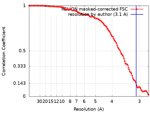
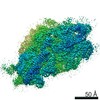
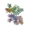
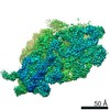
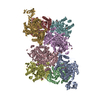
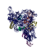
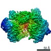
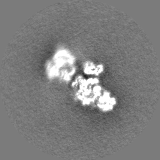
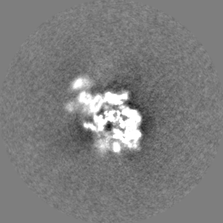
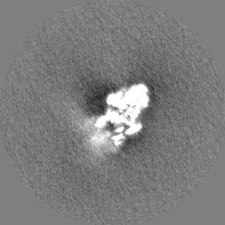
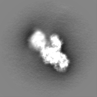
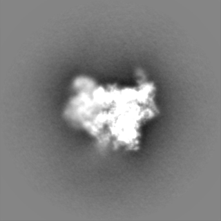
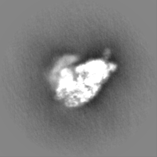
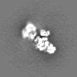
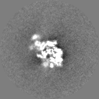
ジャーナル: Nucleic Acids Res / 年: 2021 タイトル: Structural basis of successive adenosine modifications by the conserved ribosomal methyltransferase KsgA. 著者: Niklas C Stephan / Anne B Ries / Daniel Boehringer / Nenad Ban / 要旨: Biogenesis of ribosomal subunits involves enzymatic modifications of rRNA that fine-tune functionally important regions. The universally conserved prokaryotic dimethyltransferase KsgA sequentially ...Biogenesis of ribosomal subunits involves enzymatic modifications of rRNA that fine-tune functionally important regions. The universally conserved prokaryotic dimethyltransferase KsgA sequentially modifies two universally conserved adenosine residues in helix 45 of the small ribosomal subunit rRNA, which is in proximity of the decoding site. Here we present the cryo-EM structure of Escherichia coli KsgA bound to an E. coli 30S at a resolution of 3.1 Å. The high-resolution structure reveals how KsgA recognizes immature rRNA and binds helix 45 in a conformation where one of the substrate nucleotides is flipped-out into the active site. We suggest that successive processing of two adjacent nucleotides involves base-flipping of the rRNA, which allows modification of the second substrate nucleotide without dissociation of the enzyme. Since KsgA is homologous to the essential eukaryotic methyltransferase Dim1 involved in 40S maturation, these results have also implications for understanding eukaryotic ribosome maturation.
 ムービー
ムービー コントローラー


 データを開く
 基本情報
基本情報 マップデータ
 試料
試料 キーワード
キーワード 機能・相同性情報
 データ登録者
 スイス, 1件
  引用
引用 構造の表示
構造の表示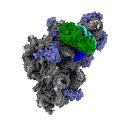
 ダウンロードとリンク
 emd_12736.png
 http://ftp.pdbj.org/pub/emdb/structures/EMD-12736


 Z (Sec.)
 Y (Row.)
 X (Col.)


 試料の構成要素
 解析
解析 電子顕微鏡法
 FIELD EMISSION GUN