ジャーナル: J Mol Biol / 年: 2006 タイトル: Structure of the dodecahedral penton particle from human adenovirus type 3. 著者: P Fuschiotti / G Schoehn / P Fender / C M S Fabry / E A Hewat / J Chroboczek / R W H Ruigrok / J F Conway / 要旨: The sub-viral dodecahedral particle of human adenovirus type 3, composed of the viral penton base and fiber proteins, shares an important characteristic of the entire virus: it can attach to cells ...The sub-viral dodecahedral particle of human adenovirus type 3, composed of the viral penton base and fiber proteins, shares an important characteristic of the entire virus: it can attach to cells and penetrate them. Structure determination of the fiberless dodecahedron by cryo-electron microscopy to 9 Angstroms resolution reveals tightly bound pentamer subunits, with only minimal interfaces between penton bases stabilizing the fragile dodecahedron. The internal cavity of the dodecahedron is approximately 80 Angstroms in diameter, and the interior surface is accessible to solvent through perforations of approximately 20 Angstroms diameter between the pentamer towers. We observe weak density beneath pentamers that we attribute to a penton base peptide including residues 38-48. The intact amino-terminal domain appears to interfere with pentamer-pentamer interactions and its absence by mutation or proteolysis is essential for dodecamer assembly. Differences between the 9 Angstroms dodecahedron structure and the adenovirus serotype 2 (Ad2) crystallographic model correlate closely with differences in sequence. The 3D structure of the dodecahedron including fibers at 16 Angstroms resolution reveals extra density on the top of the penton base that can be attributed to the fiber N terminus. The fiber itself exhibits striations that correlate with features of the atomic structure of the partial Ad2 fiber and that represent a repeat motif present in the amino acid sequence. These new observations offer important insights into particle assembly and stability, as well as the practicality of using the dodecahedron in targeted drug delivery. The structural work provides a sound basis for manipulating the properties of this particle and thereby enhancing its value for such therapeutic use.
#200 - 2016年8月 正二十面体型ウイルスの準対称性 (Quasisymmetry in Icosahedral Viruses) 類似性 (1)
-
マップ
ファイル
ダウンロード / ファイル: emd_1179.map.gz / 形式: CCP4 / 大きさ: 30.9 MB / タイプ: IMAGE STORED AS SIGNED INTEGER (2 BYTES)
注釈
The pixel size has been calibrated using the X-ray structure of adenovirus 2 penton base. Accession number 1X9P
ボクセルのサイズ
X=Y=Z: 1.72 Å
密度
表面レベル
登録者による: 79.0 / ムービー #1: 70
最小 - 最大
-357.0 - 348.0
平均 (標準偏差)
2.10371828 (±44.685005189999998)
対称性
空間群: 1
詳細
EMDB XML:
マップ形状
Axis order
X
Y
Z
Origin
-127
-127
-127
サイズ
255
255
255
Spacing
255
255
255
セル
A=B=C: 438.6 Å α=β=γ: 90.0 °
CCP4マップ ヘッダ情報:
mode
Image stored as Integer*27
Å/pix. X/Y/Z
1.72
1.72
1.72
M x/y/z
255
255
255
origin x/y/z
0.000
0.000
0.000
length x/y/z
438.600
438.600
438.600
α/β/γ
90.000
90.000
90.000
start NX/NY/NZ
-77
-77
0
NX/NY/NZ
155
155
78
MAP C/R/S
1
2
3
start NC/NR/NS
-127
-127
-127
NC/NR/NS
255
255
255
D min/max/mean
-357.000
348.000
2.104
-
添付データ
-
試料の構成要素
-
全体 : adenovirus 3 penton dodecahedron
全体
名称: adenovirus 3 penton dodecahedron
要素
試料: adenovirus 3 penton dodecahedron
タンパク質・ペプチド: Ad3 penton base
タンパク質・ペプチド: Ad3 fiber
-
超分子 #1000: adenovirus 3 penton dodecahedron
超分子
名称: adenovirus 3 penton dodecahedron / タイプ: sample / ID: 1000 詳細: The pentons were expressed in baculovirus and the penton self-assemble into dodecahedrons 集合状態: 12 pentameric penton base and 12 trimeric fiber Number unique components: 2
生物種: Human adenovirus 3 (ヒトアデノウイルス) / 別称: human adenovirus 3
分子量
実験値: 35 KDa
組換発現
生物種: bacculovirus (ウイルス) / 組換プラスミド: pAcUW31
-
実験情報
-
構造解析
手法
ネガティブ染色法, クライオ電子顕微鏡法
解析
単粒子再構成法
試料の集合状態
particle
-
試料調製
濃度
1 mg/mL
緩衝液
pH: 6.6 / 詳細: 25 mM phosphate buffer at pH 6.6
染色
タイプ: NEGATIVE 詳細: Quantifoil R2 1 grids (Quantifoil Micro Tools GmbH, Germany) were loaded with 4 ul of sample at 1 mg ml, blotted and rapidly frozen in liquid ethane within a liquid nitrogen bath using a Zeiss cryoplunger
グリッド
詳細: Quantifoil R2/1 grids
凍結
凍結剤: ETHANE / チャンバー内温度: 100 K / 装置: OTHER / 詳細: Vitrification instrument: Zeiss cryoplunger
-
電子顕微鏡法
顕微鏡
FEI/PHILIPS CM200T
温度
平均: 98 K
アライメント法
Legacy - 非点収差: objective lens astigmatism was corrected at 100,000 times magnification
 ムービー
ムービー コントローラー


 データを開く
 基本情報
基本情報 マップデータ
 試料
試料 機能・相同性情報
 Human adenovirus 3 (ヒトアデノウイルス)
 データ登録者
 引用
引用
 構造の表示
構造の表示 UCSF Chimera
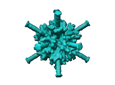
 ダウンロードとリンク
 1179.gif
 http://ftp.pdbj.org/pub/emdb/structures/EMD-1179


 試料の構成要素
 解析
解析 電子顕微鏡法