+ データを開く
- 基本情報
基本情報
| 登録情報 | データベース: EMDB / ID: EMD-10593 | |||||||||
|---|---|---|---|---|---|---|---|---|---|---|
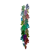
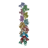
| タイトル | Cryo-EM structure of Pf4 bacteriophage coat protein with single-stranded DNA | |||||||||
 マップデータ マップデータ | ||||||||||
 試料 試料 |
| |||||||||
 キーワード キーワード | Bacteriophage / helical / filamentous / VIRUS | |||||||||
| 機能・相同性 | Inovirus Coat protein B / Capsid protein G8P / helical viral capsid / host cell membrane / membrane / Capsid protein G8P / Coat protein B of bacteriophage Pf1 機能・相同性情報 機能・相同性情報 | |||||||||
| 生物種 |  Pseudomonas virus Pf1 (ウイルス) / Pseudomonas virus Pf1 (ウイルス) /  Pseudomonas aeruginosa (strain ATCC 15692 / DSM 22644 / CIP 104116 / JCM 14847 / LMG 12228 / 1C / PRS 101 / PAO1) (緑膿菌) / Pseudomonas aeruginosa (strain ATCC 15692 / DSM 22644 / CIP 104116 / JCM 14847 / LMG 12228 / 1C / PRS 101 / PAO1) (緑膿菌) /  Pseudomonas aeruginosa PAO1 (緑膿菌) Pseudomonas aeruginosa PAO1 (緑膿菌) | |||||||||
| 手法 | らせん対称体再構成法 / クライオ電子顕微鏡法 / 解像度: 3.2 Å | |||||||||
 データ登録者 データ登録者 | Tarafder AK / von Kugelgen A | |||||||||
| 資金援助 |  英国, 1件 英国, 1件
| |||||||||
 引用 引用 |  ジャーナル: Proc Natl Acad Sci U S A / 年: 2020 ジャーナル: Proc Natl Acad Sci U S A / 年: 2020タイトル: Phage liquid crystalline droplets form occlusive sheaths that encapsulate and protect infectious rod-shaped bacteria. 著者: Abul K Tarafder / Andriko von Kügelgen / Adam J Mellul / Ulrike Schulze / Dirk G A L Aarts / Tanmay A M Bharat /  要旨: The opportunistic pathogen is a major cause of antibiotic-tolerant infections in humans. evades antibiotics in bacterial biofilms by up-regulating expression of a symbiotic filamentous inoviral ...The opportunistic pathogen is a major cause of antibiotic-tolerant infections in humans. evades antibiotics in bacterial biofilms by up-regulating expression of a symbiotic filamentous inoviral prophage, Pf4. We investigated the mechanism of phage-mediated antibiotic tolerance using biochemical reconstitution combined with structural biology and high-resolution cellular imaging. We resolved electron cryomicroscopy atomic structures of Pf4 with and without its linear single-stranded DNA genome, and studied Pf4 assembly into liquid crystalline droplets using optical microscopy and electron cryotomography. By biochemically replicating conditions necessary for antibiotic protection, we found that phage liquid crystalline droplets form phase-separated occlusive compartments around rod-shaped bacteria leading to increased bacterial survival. Encapsulation by these compartments was observed even when inanimate colloidal rods were used to mimic rod-shaped bacteria, suggesting that shape and size complementarity profoundly influences the process. Filamentous inoviruses are pervasive across prokaryotes, and in particular, several Gram-negative bacterial pathogens including , and harbor these prophages. We propose that biophysical occlusion mediated by secreted filamentous molecules such as Pf4 may be a general strategy of bacterial survival in harsh environments. | |||||||||
| 履歴 |
|
- 構造の表示
構造の表示
| ムービー |
 ムービービューア ムービービューア |
|---|---|
| 構造ビューア | EMマップ:  SurfView SurfView Molmil Molmil Jmol/JSmol Jmol/JSmol |
| 添付画像 |
- ダウンロードとリンク
-EMDBアーカイブ
| マップデータ |  emd_10593.map.gz emd_10593.map.gz | 227.1 MB |  EMDBマップデータ形式 EMDBマップデータ形式 | |
|---|---|---|---|---|
| ヘッダ (付随情報) |  emd-10593-v30.xml emd-10593-v30.xml emd-10593.xml emd-10593.xml | 17.4 KB 17.4 KB | 表示 表示 |  EMDBヘッダ EMDBヘッダ |
| 画像 |  emd_10593.png emd_10593.png | 79.1 KB | ||
| Filedesc metadata |  emd-10593.cif.gz emd-10593.cif.gz | 6.1 KB | ||
| アーカイブディレクトリ |  http://ftp.pdbj.org/pub/emdb/structures/EMD-10593 http://ftp.pdbj.org/pub/emdb/structures/EMD-10593 ftp://ftp.pdbj.org/pub/emdb/structures/EMD-10593 ftp://ftp.pdbj.org/pub/emdb/structures/EMD-10593 | HTTPS FTP |
-検証レポート
| 文書・要旨 |  emd_10593_validation.pdf.gz emd_10593_validation.pdf.gz | 514.7 KB | 表示 |  EMDB検証レポート EMDB検証レポート |
|---|---|---|---|---|
| 文書・詳細版 |  emd_10593_full_validation.pdf.gz emd_10593_full_validation.pdf.gz | 514.2 KB | 表示 | |
| XML形式データ |  emd_10593_validation.xml.gz emd_10593_validation.xml.gz | 6.9 KB | 表示 | |
| CIF形式データ |  emd_10593_validation.cif.gz emd_10593_validation.cif.gz | 8 KB | 表示 | |
| アーカイブディレクトリ |  https://ftp.pdbj.org/pub/emdb/validation_reports/EMD-10593 https://ftp.pdbj.org/pub/emdb/validation_reports/EMD-10593 ftp://ftp.pdbj.org/pub/emdb/validation_reports/EMD-10593 ftp://ftp.pdbj.org/pub/emdb/validation_reports/EMD-10593 | HTTPS FTP |
-関連構造データ
- リンク
リンク
| EMDBのページ |  EMDB (EBI/PDBe) / EMDB (EBI/PDBe) /  EMDataResource EMDataResource |
|---|---|
| 「今月の分子」の関連する項目 |
- マップ
マップ
| ファイル |  ダウンロード / ファイル: emd_10593.map.gz / 形式: CCP4 / 大きさ: 244.1 MB / タイプ: IMAGE STORED AS FLOATING POINT NUMBER (4 BYTES) ダウンロード / ファイル: emd_10593.map.gz / 形式: CCP4 / 大きさ: 244.1 MB / タイプ: IMAGE STORED AS FLOATING POINT NUMBER (4 BYTES) | ||||||||||||||||||||||||||||||||||||||||||||||||||||||||||||
|---|---|---|---|---|---|---|---|---|---|---|---|---|---|---|---|---|---|---|---|---|---|---|---|---|---|---|---|---|---|---|---|---|---|---|---|---|---|---|---|---|---|---|---|---|---|---|---|---|---|---|---|---|---|---|---|---|---|---|---|---|---|
| 投影像・断面図 | 画像のコントロール
画像は Spider により作成 | ||||||||||||||||||||||||||||||||||||||||||||||||||||||||||||
| ボクセルのサイズ | X=Y=Z: 1.38 Å | ||||||||||||||||||||||||||||||||||||||||||||||||||||||||||||
| 密度 |
| ||||||||||||||||||||||||||||||||||||||||||||||||||||||||||||
| 対称性 | 空間群: 1 | ||||||||||||||||||||||||||||||||||||||||||||||||||||||||||||
| 詳細 | EMDB XML:
CCP4マップ ヘッダ情報:
| ||||||||||||||||||||||||||||||||||||||||||||||||||||||||||||
-添付データ
- 試料の構成要素
-全体 : Pseudomonas virus Pf1
| 全体 | 名称:  Pseudomonas virus Pf1 (ウイルス) Pseudomonas virus Pf1 (ウイルス) |
|---|---|
| 要素 |
|
-超分子 #1: Pseudomonas virus Pf1
| 超分子 | 名称: Pseudomonas virus Pf1 / タイプ: virus / ID: 1 / 親要素: 0 / 含まれる分子: all / ウイルスタイプ: VIRION / ウイルス・単離状態: STRAIN / ウイルス・エンベロープ: No / ウイルス・中空状態: No |
|---|---|
| 宿主 | 生物種:  Pseudomonas aeruginosa (strain ATCC 15692 / DSM 22644 / CIP 104116 / JCM 14847 / LMG 12228 / 1C / PRS 101 / PAO1) (緑膿菌) Pseudomonas aeruginosa (strain ATCC 15692 / DSM 22644 / CIP 104116 / JCM 14847 / LMG 12228 / 1C / PRS 101 / PAO1) (緑膿菌) |
| ウイルス殻 | Shell ID: 1 / 名称: Coat protein B (CoaB) / 直径: 62.0 Å |
-超分子 #2: Pseudomonas virus Pf1
| 超分子 | 名称: Pseudomonas virus Pf1 / タイプ: complex / ID: 2 / 親要素: 1 / 含まれる分子: #1 |
|---|---|
| 由来(天然) | 生物種:  Pseudomonas virus Pf1 (ウイルス) Pseudomonas virus Pf1 (ウイルス) |
-超分子 #3: DNA
| 超分子 | 名称: DNA / タイプ: complex / ID: 3 / 親要素: 1 / 含まれる分子: #2 |
|---|---|
| 由来(天然) | 生物種:  Pseudomonas aeruginosa (strain ATCC 15692 / DSM 22644 / CIP 104116 / JCM 14847 / LMG 12228 / 1C / PRS 101 / PAO1) (緑膿菌) Pseudomonas aeruginosa (strain ATCC 15692 / DSM 22644 / CIP 104116 / JCM 14847 / LMG 12228 / 1C / PRS 101 / PAO1) (緑膿菌) |
-分子 #1: Coat protein B of bacteriophage Pf1
| 分子 | 名称: Coat protein B of bacteriophage Pf1 / タイプ: protein_or_peptide / ID: 1 / コピー数: 1 / 光学異性体: LEVO |
|---|---|
| 由来(天然) | 生物種:  Pseudomonas virus Pf1 (ウイルス) Pseudomonas virus Pf1 (ウイルス) |
| 分子量 | 理論値: 4.612393 KDa |
| 配列 | 文字列: GVIDTSAVES AITDGQGDMK AIGGYIVGAL VILAVAGLIY SMLRKA UniProtKB: Coat protein B of bacteriophage Pf1 |
-分子 #2: DNA (5'-D(P*AP*AP*AP*AP*AP*A)-3')
| 分子 | 名称: DNA (5'-D(P*AP*AP*AP*AP*AP*A)-3') / タイプ: dna / ID: 2 / 詳細: Model of pf4 single-stranded DNA genome / コピー数: 1 / 分類: DNA |
|---|---|
| 由来(天然) | 生物種:  Pseudomonas aeruginosa PAO1 (緑膿菌) Pseudomonas aeruginosa PAO1 (緑膿菌) |
| 分子量 | 理論値: 1.834283 KDa |
| 配列 | 文字列: (DA)(DA)(DA)(DA)(DA)(DA) |
-実験情報
-構造解析
| 手法 | クライオ電子顕微鏡法 |
|---|---|
 解析 解析 | らせん対称体再構成法 |
| 試料の集合状態 | filament |
- 試料調製
試料調製
| 濃度 | 5 mg/mL | |||||||||||||||
|---|---|---|---|---|---|---|---|---|---|---|---|---|---|---|---|---|
| 緩衝液 | pH: 7.4 構成要素:
詳細: 1x Phosphate buffered saline | |||||||||||||||
| グリッド | モデル: Quantifoil R2/2 / 材質: COPPER/RHODIUM / メッシュ: 200 / 支持フィルム - 材質: CARBON / 支持フィルム - トポロジー: HOLEY / 前処理 - タイプ: GLOW DISCHARGE / 前処理 - 時間: 20 sec. / 前処理 - 雰囲気: AIR / 前処理 - 気圧: 0.03 kPa 詳細: 20 second glow discharge at 15 mA in a LeicaEM ACE200 | |||||||||||||||
| 凍結 | 凍結剤: ETHANE / チャンバー内湿度: 100 % / チャンバー内温度: 283 K / 装置: FEI VITROBOT MARK IV 詳細: Samples for cryo-EM were prepared by pipetting 2.5 ul of the sample onto freshly glow-discharged Quantifoil grids (Cu/Rh R2/2, 200 mesh). Grids were blotted for 2.5 seconds with a blot force ...詳細: Samples for cryo-EM were prepared by pipetting 2.5 ul of the sample onto freshly glow-discharged Quantifoil grids (Cu/Rh R2/2, 200 mesh). Grids were blotted for 2.5 seconds with a blot force of -15, 0.5 second drain and 0 second wait times.. | |||||||||||||||
| 詳細 | Filamentous phage purified from source |
- 電子顕微鏡法
| 顕微鏡 | FEI TITAN KRIOS |
|---|---|
| 温度 | 最低: 80.0 K / 最高: 80.0 K |
| 特殊光学系 | エネルギーフィルター - 名称: GIF Bioquantum / エネルギーフィルター - スリット幅: 20 eV |
| 撮影 | フィルム・検出器のモデル: GATAN K2 SUMMIT (4k x 4k) 検出モード: COUNTING / デジタル化 - 画像ごとのフレーム数: 1-40 / 撮影したグリッド数: 2 / 実像数: 4110 / 平均露光時間: 10.0 sec. / 平均電子線量: 43.0 e/Å2 |
| 電子線 | 加速電圧: 300 kV / 電子線源:  FIELD EMISSION GUN FIELD EMISSION GUN |
| 電子光学系 | C2レンズ絞り径: 50.0 µm / 最大 デフォーカス(補正後): -3.0 µm / 最小 デフォーカス(補正後): -1.0 µm / 倍率(補正後): 105000 / 照射モード: FLOOD BEAM / 撮影モード: BRIGHT FIELD / Cs: 2.7 mm / 最大 デフォーカス(公称値): -3.0 µm / 最小 デフォーカス(公称値): -1.0 µm / 倍率(公称値): 105000 |
| 試料ステージ | 試料ホルダーモデル: FEI TITAN KRIOS AUTOGRID HOLDER ホルダー冷却材: NITROGEN |
| 実験機器 |  モデル: Titan Krios / 画像提供: FEI Company |
- 画像解析
画像解析
| 最終 再構成 | 使用したクラス数: 1 想定した対称性 - らせんパラメータ - Δz: 3.14 Å 想定した対称性 - らせんパラメータ - ΔΦ: 65.9 ° 想定した対称性 - らせんパラメータ - 軸対称性: C1 (非対称) アルゴリズム: FOURIER SPACE / 解像度のタイプ: BY AUTHOR / 解像度: 3.2 Å / 解像度の算出法: FSC 0.143 CUT-OFF / ソフトウェア - 名称: RELION (ver. 3.0) / 使用した粒子像数: 185002 |
|---|---|
| Segment selection | 選択した数: 351381 / ソフトウェア - 名称: RELION (ver. 2.0) |
| 初期モデル | モデルのタイプ: NONE / 詳細: Fourier-Bessel indexing |
| 最終 角度割当 | タイプ: NOT APPLICABLE / ソフトウェア - 名称: RELION (ver. 2.0) |
-原子モデル構築 1
| 詳細 | The helical filament symmetry is only applied to the Pf4 coat protein and not the DNA. The reason for this is that the DNA bases are not resolved clearly due to averaging along the ssDNA genome of the phage, meaning that refinement of the protein into the map is of a higher quality than the ssDNA, where a poly-adenine model has been built. |
|---|---|
| 精密化 | 空間: REAL / プロトコル: BACKBONE TRACE / 温度因子: 93.74 / 当てはまり具合の基準: Correlation coefficient |
| 得られたモデル |  PDB-6tup: |
 ムービー
ムービー コントローラー


 Z (Sec.)
 Y (Row.)
 X (Col.)


