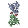
 Movie
Movie Controller


+ Open data

- Basic information
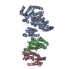
| Entry | Database: EMDB / ID: EMD-0924 | |||||||||
|---|---|---|---|---|---|---|---|---|---|---|
| Title | mutation transporter state2-1 | |||||||||
 Map data Map data | sharpened map | |||||||||
 Sample Sample |
| |||||||||
 Keywords Keywords | calcium / METAL TRANSPORT | |||||||||
| Function / homology |  Function and homology information Function and homology informationER-nucleus signaling pathway / longitudinal sarcoplasmic reticulum / positive regulation of endoplasmic reticulum calcium ion concentration / P-type calcium transporter activity involved in regulation of cardiac muscle cell membrane potential / calcium ion transport from cytosol to endoplasmic reticulum / regulation of calcium ion-dependent exocytosis of neurotransmitter / calcium ion-transporting ATPase complex / T-tubule organization / regulation of cardiac muscle cell action potential involved in regulation of contraction / regulation of cardiac muscle cell membrane potential ...ER-nucleus signaling pathway / longitudinal sarcoplasmic reticulum / positive regulation of endoplasmic reticulum calcium ion concentration / P-type calcium transporter activity involved in regulation of cardiac muscle cell membrane potential / calcium ion transport from cytosol to endoplasmic reticulum / regulation of calcium ion-dependent exocytosis of neurotransmitter / calcium ion-transporting ATPase complex / T-tubule organization / regulation of cardiac muscle cell action potential involved in regulation of contraction / regulation of cardiac muscle cell membrane potential / calcium ion import into sarcoplasmic reticulum / platelet dense tubular network membrane / Pre-NOTCH Processing in Golgi / sarcoplasmic reticulum calcium ion transport / ribbon synapse / P-type Ca2+ transporter / negative regulation of heart contraction / P-type calcium transporter activity / transition between fast and slow fiber / cardiac muscle hypertrophy in response to stress / regulation of the force of heart contraction / endoplasmic reticulum calcium ion homeostasis / S100 protein binding / relaxation of cardiac muscle / regulation of cardiac muscle contraction by calcium ion signaling / Reduction of cytosolic Ca++ levels / organelle localization by membrane tethering / : / autophagosome membrane docking / lncRNA binding / epidermis development / Ion transport by P-type ATPases / regulation of cardiac conduction / autophagosome assembly / positive regulation of heart rate / Ion homeostasis / positive regulation of cardiac muscle cell apoptotic process / sarcoplasmic reticulum membrane / response to endoplasmic reticulum stress / sarcoplasmic reticulum / neuron cellular homeostasis / calcium channel regulator activity / intracellular calcium ion homeostasis / calcium ion transmembrane transport / cellular response to oxidative stress / monoatomic ion transmembrane transport / transmembrane transporter binding / cell adhesion / calcium ion binding / endoplasmic reticulum membrane / enzyme binding / endoplasmic reticulum / ATP hydrolysis activity / ATP binding / membrane / plasma membrane Similarity search - Function | |||||||||
| Biological species |  Homo sapiens (human) Homo sapiens (human) | |||||||||
| Method | single particle reconstruction / cryo EM / Resolution: 2.8 Å | |||||||||
 Authors Authors | Zhang Y / Tsutsumi A | |||||||||
| Funding support |  Japan, 1 items Japan, 1 items
| |||||||||
 Citation Citation | Journal: Sci Adv / Year: 2020 Title: Cryo-EM structures of SERCA2b reveal the mechanism of regulation by the luminal extension tail. Authors: Yuxia Zhang / Michio Inoue / Akihisa Tsutsumi / Satoshi Watanabe / Tomohiro Nishizawa / Kazuhiro Nagata / Masahide Kikkawa / Kenji Inaba / Abstract: Sarco/endoplasmic reticulum Ca ATPase (SERCA) pumps Ca from the cytosol into the ER and maintains the cellular calcium homeostasis. Herein, we present cryo-electron microscopy (cryo-EM) structures of ...Sarco/endoplasmic reticulum Ca ATPase (SERCA) pumps Ca from the cytosol into the ER and maintains the cellular calcium homeostasis. Herein, we present cryo-electron microscopy (cryo-EM) structures of human SERCA2b in E1∙2Ca-adenylyl methylenediphosphonate (AMPPCP) and E2-BeF states at 2.9- and 2.8-Å resolutions, respectively. The structures revealed that the luminal extension tail (LE) characteristic of SERCA2b runs parallel to the lipid-water boundary near the luminal ends of transmembrane (TM) helices TM10 and TM7 and approaches the luminal loop flanked by TM7 and TM8. While the LE served to stabilize the cytosolic and TM domain arrangement of SERCA2b, deletion of the LE rendered the overall conformation resemble that of SERCA1a and SERCA2a and allowed multiple conformations. Thus, the LE appears to play a critical role in conformational regulation in SERCA2b, which likely explains the different kinetic properties of SERCA2b from those of other isoforms lacking the LE. | |||||||||
| History |
|
- Structure visualization
| Movie |
Movie viewer |
|---|---|
| Structure viewer | EM map: SurfViewMolmilJmol/JSmol |
| Supplemental images |

- Downloads & links
-EMDB archive
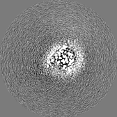
| Map data | emd_0924.map.gz | 2.7 MB | EMDB map data format | |
|---|---|---|---|---|
| Header (meta data) | emd-0924-v30.xmlemd-0924.xml | 22.1 KB 22.1 KB | Display Display | EMDB header |
| FSC (resolution estimation) | emd_0924_fsc.xml | 6 KB | Display | FSC data file |
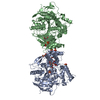
| Images | 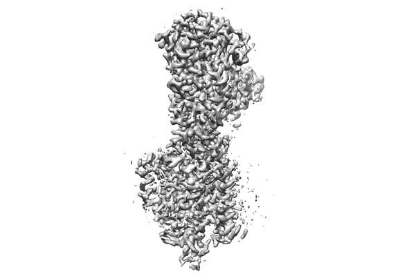 emd_0924.png emd_0924.png | 44.7 KB | ||
| Masks | emd_0924_msk_1.map | 18.1 MB | Mask map | |
| Filedesc metadata | emd-0924.cif.gz | 7 KB | ||
| Others | emd_0924_additional.map.gzemd_0924_half_map_1.map.gzemd_0924_half_map_2.map.gz | 13.7 MB 13.8 MB 13.8 MB | ||
| Archive directory |  http://ftp.pdbj.org/pub/emdb/structures/EMD-0924ftp://ftp.pdbj.org/pub/emdb/structures/EMD-0924 http://ftp.pdbj.org/pub/emdb/structures/EMD-0924ftp://ftp.pdbj.org/pub/emdb/structures/EMD-0924 | HTTPS FTP |
-Related structure data
| Related structure data |  6ln5MC 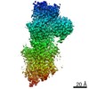 0912C  0915C 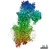 0925C 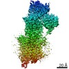 0926C 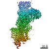 0927C 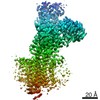 0928C 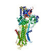 6lleC 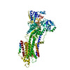 6llyC 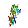 6ln6C 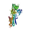 6ln7C 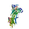 6ln8C 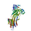 6ln9C M: atomic model generated by this map C: citing same article ( |
|---|---|
| Similar structure data |


-Links
| EMDB pages | EMDB (EBI/PDBe) / EMDataResource |
|---|---|
| Related items in Molecule of the Month |


-Map
| File | Download / File: emd_0924.map.gz / Format: CCP4 / Size: 18.1 MB / Type: IMAGE STORED AS FLOATING POINT NUMBER (4 BYTES) | ||||||||||||||||||||||||||||||||||||||||||||||||||||||||||||
|---|---|---|---|---|---|---|---|---|---|---|---|---|---|---|---|---|---|---|---|---|---|---|---|---|---|---|---|---|---|---|---|---|---|---|---|---|---|---|---|---|---|---|---|---|---|---|---|---|---|---|---|---|---|---|---|---|---|---|---|---|---|
| Annotation | sharpened map | ||||||||||||||||||||||||||||||||||||||||||||||||||||||||||||
| Projections & slices | Image control
Images are generated by Spider. | ||||||||||||||||||||||||||||||||||||||||||||||||||||||||||||
| Voxel size | X=Y=Z: 1.245 Å | ||||||||||||||||||||||||||||||||||||||||||||||||||||||||||||
| Density |
| ||||||||||||||||||||||||||||||||||||||||||||||||||||||||||||
| Symmetry | Space group: 1 | ||||||||||||||||||||||||||||||||||||||||||||||||||||||||||||
| Details | EMDB XML:
CCP4 map header:
| ||||||||||||||||||||||||||||||||||||||||||||||||||||||||||||
 Z (Sec.)
 Y (Row.)
 X (Col.)


-Supplemental data
-Mask #1
| File | emd_0924_msk_1.map | ||||||||||||
|---|---|---|---|---|---|---|---|---|---|---|---|---|---|
| Projections & Slices |
| ||||||||||||
| Density Histograms |


-Additional map: mono-sharpened map
| File | emd_0924_additional.map | ||||||||||||
|---|---|---|---|---|---|---|---|---|---|---|---|---|---|
| Annotation | mono-sharpened map | ||||||||||||
| Projections & Slices |
| ||||||||||||
| Density Histograms |


-Half map: #1
| File | emd_0924_half_map_1.map | ||||||||||||
|---|---|---|---|---|---|---|---|---|---|---|---|---|---|
| Projections & Slices |
| ||||||||||||
| Density Histograms |


-Half map: #2
| File | emd_0924_half_map_2.map | ||||||||||||
|---|---|---|---|---|---|---|---|---|---|---|---|---|---|
| Projections & Slices |
| ||||||||||||
| Density Histograms |


- Sample components
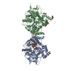
-Entire : SERCA2b MUTATION with AMPPCP -1
| Entire | Name: SERCA2b MUTATION with AMPPCP -1 |
|---|---|
| Components |
|
-Supramolecule #1: SERCA2b MUTATION with AMPPCP -1
| Supramolecule | Name: SERCA2b MUTATION with AMPPCP -1 / type: complex / ID: 1 / Parent: 0 / Macromolecule list: #1 |
|---|---|
| Source (natural) | Organism: Homo sapiens (human) |
| Molecular weight | Theoretical: 110 kDa/nm |
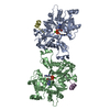
-Macromolecule #1: Sarcoplasmic/endoplasmic reticulum calcium ATPase 2
| Macromolecule | Name: Sarcoplasmic/endoplasmic reticulum calcium ATPase 2 / type: protein_or_peptide / ID: 1 / Number of copies: 1 / Enantiomer: LEVO / EC number: P-type Ca2+ transporter |
|---|---|
| Source (natural) | Organism: Homo sapiens (human) |
| Molecular weight | Theoretical: 116.432453 KDa |
| Recombinant expression | Organism: Homo sapiens (human) |
| Sequence | String: MGGVAMPGAE DDVVRENLYF QGKDGLAAME NAHTKTVEEV LGHFGVNEST GLSLEQVKKL KERWGSNELP AEEGKTLLEL VIEQFEDLL VRILLLAACI SFVLAWFEEG EETITAFVEP FVILLILVAN AIVGVWQERN AENAIEALKE YEPEMGKVYR Q DRKSVQRI ...String: MGGVAMPGAE DDVVRENLYF QGKDGLAAME NAHTKTVEEV LGHFGVNEST GLSLEQVKKL KERWGSNELP AEEGKTLLEL VIEQFEDLL VRILLLAACI SFVLAWFEEG EETITAFVEP FVILLILVAN AIVGVWQERN AENAIEALKE YEPEMGKVYR Q DRKSVQRI KAKDIVPGDI VEIAVGDKVP ADIRLTSIKS TTLRVDQSIL TGESVSVIKH TDPVPDPRAV NQDKKNMLFS GT NIAAGKA MGVVVATGVN TEIGKIRDEM VATEQERTPL QQKLDEFGEQ LSKVISLICI AVWIINIGHF NDPVHGGSWI RGA IYYFKI AVALAVAAIP EGLPAVITTC LALGTRRMAK KNAIVRSLPS VETLGCTSVI CSDKTGTLTT NQMSVCRMFI LDRV EGDTC SLNEFTITGS TYAPIGEVHK DDKPVNCHQY DGLVELATIC ALCNDSALDY NEAKGVYEKV GEATETALTC LVEKM NVFD TELKGLSKIE RANACNSVIK QLMKKEFTLE FSRDRKSMSV YCTPNKPSRT SMSKMFVKGA PEGVIDRCTH IRVGST KVP MTSGVKQKIM SVIREWGSGS DTLRCLALAT HDNPLRREEM HLEDSANFIK YETNLTFVGC VGMLDPPRIE VASSVKL CR QAGIRVIMIT GDNKGTAVAI CRRIGIFGQD EDVTSKAFTG REFDELNPSA QRDACLNARC FARVEPSHKS KIVEFLQS F DEITAMTGDG VNDAPALKKA EIGIAMGSGT AVAKTASEMV LADDNFSTIV AAVEEGRAIY NNMKQFIRYL ISSNVGEVV CIFLTAALGF PEALIPVQLL WVNLVTDGLP ATALGFNPPD LDIMNKPPRN PKEPLISGWL FFRYLAIGCY VGAATVGAAA WWFIAADGG PRVSFYQLSH FLQCKEDNPD FEGVDCAIFE SPYPMTMALS VLVTIEMCNA LNSLSENQSL LRMPPWENIW L VGSICLSM SLHFLILYVE PLPLIFQITP LNVTQWLMVL KISLPVILMD ETLKFVARNY LEPGKECVQP ATKSCSFSAC TD GISWPFV LLIMPLVIWV YS UniProtKB: Sarcoplasmic/endoplasmic reticulum calcium ATPase 2 |
-Macromolecule #2: PHOSPHOMETHYLPHOSPHONIC ACID ADENYLATE ESTER
| Macromolecule | Name: PHOSPHOMETHYLPHOSPHONIC ACID ADENYLATE ESTER / type: ligand / ID: 2 / Number of copies: 1 / Formula: ACP |
|---|---|
| Molecular weight | Theoretical: 505.208 Da |
| Chemical component information |  ChemComp-ACP: |
-Macromolecule #3: MAGNESIUM ION
| Macromolecule | Name: MAGNESIUM ION / type: ligand / ID: 3 / Number of copies: 1 / Formula: MG |
|---|---|
| Molecular weight | Theoretical: 24.305 Da |
-Macromolecule #4: CALCIUM ION
| Macromolecule | Name: CALCIUM ION / type: ligand / ID: 4 / Number of copies: 2 / Formula: CA |
|---|---|
| Molecular weight | Theoretical: 40.078 Da |
-Experimental details
-Structure determination
| Method | cryo EM |
|---|---|
 Processing Processing | single particle reconstruction |
| Aggregation state | particle |
-Sample preparation
| Buffer | pH: 7 |
|---|---|
| Vitrification | Cryogen name: ETHANE |
- Electron microscopy
| Microscope | FEI TITAN KRIOS |
|---|---|
| Image recording | Film or detector model: GATAN K3 BIOQUANTUM (6k x 4k) / Average electron dose: 60.0 e/Å2 |
| Electron beam | Acceleration voltage: 300 kV / Electron source:  FIELD EMISSION GUN FIELD EMISSION GUN |
| Electron optics | Illumination mode: OTHER / Imaging mode: BRIGHT FIELD |
| Experimental equipment |  Model: Titan Krios / Image courtesy: FEI Company |

