 Movie
Movie Controller

[English] 日本語
 Yorodumi
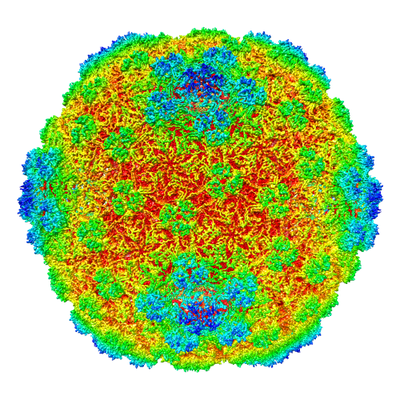
- EMDB-0618: Icosahedral reconstruction of the thermophilic bacteriophage P74-... -
+ Open data

- Basic information
| Entry | Database: EMDB / ID: EMD-0618 | |||||||||
|---|---|---|---|---|---|---|---|---|---|---|
| Title | Icosahedral reconstruction of the thermophilic bacteriophage P74-26 capsid | |||||||||
 Map data Map data | Icosahedral reconstruction of the thermophilic bacteriophage P74-26 capsid | |||||||||
 Sample Sample |
| |||||||||
 Keywords Keywords | Virus / Virion / Capsid / Major Capsid Protein / Decoration Protein / Icosahedral Virus / Caudovirus / Thermophilic / Bacteriophage | |||||||||
| Function / homology | Major capsid protein GpE / Phage major capsid protein E / viral capsid / Uncharacterized protein / Major head protein Function and homology information Function and homology information | |||||||||
| Biological species |  Thermus virus P74-26 / Thermus virus P74-26 /  Thermus phage P7426 (virus) Thermus phage P7426 (virus) | |||||||||
| Method | single particle reconstruction / cryo EM / Resolution: 2.8 Å | |||||||||
 Authors Authors | Stone NP / Demo G | |||||||||
| Funding support |  United States, 1 items United States, 1 items
| |||||||||
 Citation Citation | Journal: Nat Commun / Year: 2019 Title: Principles for enhancing virus capsid capacity and stability from a thermophilic virus capsid structure. Authors: Nicholas P Stone / Gabriel Demo / Emily Agnello / Brian A Kelch / Abstract: The capsids of double-stranded DNA viruses protect the viral genome from the harsh extracellular environment, while maintaining stability against the high internal pressure of packaged DNA. To ...The capsids of double-stranded DNA viruses protect the viral genome from the harsh extracellular environment, while maintaining stability against the high internal pressure of packaged DNA. To elucidate how capsids maintain stability in an extreme environment, we use cryoelectron microscopy to determine the capsid structure of thermostable phage P74-26 to 2.8-Å resolution. We find P74-26 capsids exhibit an overall architecture very similar to those of other tailed bacteriophages, allowing us to directly compare structures to derive the structural basis for enhanced stability. Our structure reveals lasso-like interactions that appear to function like catch bonds. This architecture allows the capsid to expand during genome packaging, yet maintain structural stability. The P74-26 capsid has T = 7 geometry despite being twice as large as mesophilic homologs. Capsid capacity is increased with a larger, flatter major capsid protein. Given these results, we predict decreased icosahedral complexity (i.e. T ≤ 7) leads to a more stable capsid assembly. | |||||||||
| History |
|
- Structure visualization
| Movie |
Movie viewer |
|---|---|
| Structure viewer | EM map: SurfViewMolmilJmol/JSmol |
| Supplemental images |
- Downloads & links
-EMDB archive
| Map data | emd_0618.map.gz | 3.5 GB | EMDB map data format | |
|---|---|---|---|---|
| Header (meta data) | emd-0618-v30.xmlemd-0618.xml | 14.9 KB 14.9 KB | Display Display | EMDB header |
| Images |  emd_0618.png emd_0618.png | 262.3 KB | ||
| Filedesc metadata | emd-0618.cif.gz | 5.9 KB | ||
| Archive directory |  http://ftp.pdbj.org/pub/emdb/structures/EMD-0618ftp://ftp.pdbj.org/pub/emdb/structures/EMD-0618 http://ftp.pdbj.org/pub/emdb/structures/EMD-0618ftp://ftp.pdbj.org/pub/emdb/structures/EMD-0618 | HTTPS FTP |
-Related structure data
| Related structure data |  6o3hMC M: atomic model generated by this map C: citing same article ( |
|---|---|
| Similar structure data |
-Links
| EMDB pages | EMDB (EBI/PDBe) / EMDataResource |
|---|
-Map
| File | Download / File: emd_0618.map.gz / Format: CCP4 / Size: 4 GB / Type: IMAGE STORED AS FLOATING POINT NUMBER (4 BYTES) | ||||||||||||||||||||||||||||||||||||||||||||||||||||||||||||
|---|---|---|---|---|---|---|---|---|---|---|---|---|---|---|---|---|---|---|---|---|---|---|---|---|---|---|---|---|---|---|---|---|---|---|---|---|---|---|---|---|---|---|---|---|---|---|---|---|---|---|---|---|---|---|---|---|---|---|---|---|---|
| Annotation | Icosahedral reconstruction of the thermophilic bacteriophage P74-26 capsid | ||||||||||||||||||||||||||||||||||||||||||||||||||||||||||||
| Projections & slices | Image control
Images are generated by Spider. | ||||||||||||||||||||||||||||||||||||||||||||||||||||||||||||
| Voxel size | X=Y=Z: 1.059 Å | ||||||||||||||||||||||||||||||||||||||||||||||||||||||||||||
| Density |
| ||||||||||||||||||||||||||||||||||||||||||||||||||||||||||||
| Symmetry | Space group: 1 | ||||||||||||||||||||||||||||||||||||||||||||||||||||||||||||
| Details | EMDB XML:
CCP4 map header:
| ||||||||||||||||||||||||||||||||||||||||||||||||||||||||||||
 Z (Sec.)
 Y (Row.)
 X (Col.)


-Supplemental data
- Sample components
-Entire : Thermus phage P7426
| Entire | Name: Thermus phage P7426 (virus) |
|---|---|
| Components |
|
-Supramolecule #1: Thermus phage P7426
| Supramolecule | Name: Thermus phage P7426 / type: virus / ID: 1 / Parent: 0 / Macromolecule list: all / NCBI-ID: 466052 / Sci species name: Thermus phage P7426 / Virus type: VIRION / Virus isolate: OTHER / Virus enveloped: No / Virus empty: No |
|---|---|
| Host (natural) | Organism:  Thermus thermophilus (bacteria) / Strain: HB8 Thermus thermophilus (bacteria) / Strain: HB8 |
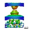
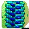
| Virus shell | Shell ID: 1 / Name: P74-26 capsid / Diameter: 824.0 Å / T number (triangulation number): 7 |
-Macromolecule #1: Major head protein
| Macromolecule | Name: Major head protein / type: protein_or_peptide / ID: 1 / Number of copies: 7 / Enantiomer: LEVO |
|---|---|
| Source (natural) | Organism: Thermus virus P74-26 |
| Molecular weight | Theoretical: 46.680754 KDa |
| Sequence | String: MRVPININNA LARVRDPLSI GGLKFPTTKE IQEAVAAIAD KFNQENDLVD RFFPEDSTFA SELELYLLRT QDAEQTGMTF VHQVGSTSL PVEARVAKVD LAKATWSPLA FKESRVWDEK EILYLGRLAD EVQAGVINEQ IAESLTWLMA RMRNRRRWLT W QVMRTGRI ...String: MRVPININNA LARVRDPLSI GGLKFPTTKE IQEAVAAIAD KFNQENDLVD RFFPEDSTFA SELELYLLRT QDAEQTGMTF VHQVGSTSL PVEARVAKVD LAKATWSPLA FKESRVWDEK EILYLGRLAD EVQAGVINEQ IAESLTWLMA RMRNRRRWLT W QVMRTGRI TIQPNDPYNP NGLKYVIDYG VTDIELPLPQ KFDAKDGNGN SAVDPIQYFR DLIKAATYFP DRRPVAIIVG PG FDEVLAD NTFVQKYVEY EKGWVVGQNT VQPPREVYRQ AALDIFKRYT GLEVMVYDKT YRDQDGSVKY WIPVGELIVL NQS TGPVGR FVYTAHVAGQ RNGKVVYATG PYLTVKDHLQ DDPPYYAIIA GFHGLPQLSG YNTEDFSFHR FKWLKYANNV QSYL PPFPP KVEL UniProtKB: Major head protein |
-Macromolecule #2: P74-26 Head Decoration Protein
| Macromolecule | Name: P74-26 Head Decoration Protein / type: protein_or_peptide / ID: 2 / Number of copies: 7 / Enantiomer: LEVO |
|---|---|
| Source (natural) | Organism: Thermus virus P74-26 |
| Molecular weight | Theoretical: 16.37663 KDa |
| Sequence | String: MDKIQLFRTI GRVQYWERVP RLHAYGVFAL PFPMDPDVEW GNWFAGPHPK AFLVSVHPSG PKAGHVYPTD LSDPDSVANV IGMVLDGHD YEADHNVTVT LRAAVPIEYV QQGIEAPPLQ PDPAVLNAAP QLKLKVIKGH YFFDYTR UniProtKB: Uncharacterized protein |
-Experimental details
-Structure determination
| Method | cryo EM |
|---|---|
 Processing Processing | single particle reconstruction |
| Aggregation state | particle |
-Sample preparation
| Buffer | pH: 8 Component:
| ||||||||||||
|---|---|---|---|---|---|---|---|---|---|---|---|---|---|
| Grid | Model: Homemade / Material: COPPER / Mesh: 400 / Support film - Material: CARBON / Support film - topology: LACEY / Pretreatment - Type: GLOW DISCHARGE / Pretreatment - Time: 45 sec. / Details: 45 second glow discharge, negative polarity, 20 mA | ||||||||||||
| Vitrification | Cryogen name: ETHANE / Chamber humidity: 95 % / Chamber temperature: 283 K / Instrument: FEI VITROBOT MARK IV |
- Electron microscopy
| Microscope | FEI TITAN KRIOS |
|---|---|
| Image recording | Film or detector model: GATAN K2 SUMMIT (4k x 4k) / Detector mode: SUPER-RESOLUTION / Number grids imaged: 2 / Number real images: 4611 / Average electron dose: 48.0 e/Å2 |
| Electron beam | Acceleration voltage: 300 kV / Electron source:  FIELD EMISSION GUN FIELD EMISSION GUN |
| Electron optics | Illumination mode: FLOOD BEAM / Imaging mode: BRIGHT FIELD / Cs: 2.7 mm / Nominal defocus max: 1.2 µm / Nominal defocus min: 0.2 µm / Nominal magnification: 130000 |
| Sample stage | Specimen holder model: FEI TITAN KRIOS AUTOGRID HOLDER / Cooling holder cryogen: NITROGEN |
| Experimental equipment |  Model: Titan Krios / Image courtesy: FEI Company |
+Image processing
-Atomic model buiding 1
| Refinement | Space: REAL / Protocol: AB INITIO MODEL / Target criteria: Correlation Coefficient |
|---|---|
| Output model | PDB-6o3h: |
