 Movie
Movie Controller
 Structure viewers
 About Yorodumi Papers


+Search query
-Structure paper
| Title | Structural insights into the mechanism of activation and inhibition of the prostaglandin D2 receptor 1. |
|---|---|
| Journal, issue, pages | Nat Commun, Vol. 16, Issue 1, Page 8944, Year 2025 |
| Publish date | Oct 8, 2025 |
 Authors Authors | Behnaz Davoudinasab / Aleksey Raskovalov / Woojin Lee / Donggyun Kim / Heesoo Kim / Jordy Homing Lam / Gye Won Han / Vsevolod Katritch / Vadim Cherezov /  |
| PubMed Abstract | The prostaglandin D2 receptor 1 (DP1), a member of the prostanoid G protein-coupled receptor (GPCR) family, plays critical roles in allergic responses, sleep regulation, immune modulation, and ...The prostaglandin D2 receptor 1 (DP1), a member of the prostanoid G protein-coupled receptor (GPCR) family, plays critical roles in allergic responses, sleep regulation, immune modulation, and vasodilation. Here, we present five high-resolution cryo-electron microscopy (cryo-EM) structures of the human DP1 receptor, including an apo structure, two inactive state structures bound to two different inverse agonists developed by ONO Pharmaceutical, and two active state structures in complex with the G protein and bound to the endogenous agonist PGD2 and its selective derivative BW245C. Structural analysis, complemented by molecular dynamics simulations and site-directed mutagenesis, reveals key residues involved in ligand recognition and suggests a distinct activation mechanism for DP1, which lacks most of the conserved class A GPCR motifs. Notably, the unique residue K76 within the conserved sodium pocket acts as a major activation switch, while amphiphilic helix 8 adopts an unconventional orientation essential for receptor function. These findings offer valuable insights into the structure and function of prostanoid receptors and may facilitate the development of therapeutics targeting DP1. |
 External links External links | Nat Commun / PubMed:41062467 / PubMed Central |
| Methods | EM (single particle) |
| Resolution | 2.41 - 3.2 Å |
| Structure data | EMDB-43839, PDB-9au0: EMDB-47802, PDB-9e9s: EMDB-47950, PDB-9ee5: EMDB-48077, PDB-9ei5: EMDB-48122, PDB-9ekh:  EMDB-71379: Cryo-EM structure of the ONO2550289-bound prostaglandin D2 receptor (DP1)-bRIL-Fab complex (Receptor-focused)  EMDB-71392: Cryo-EM structure of the ONO2550289-bound prostaglandin D2 receptor (DP1)-bRIL-Fab complex (Concensus map)  EMDB-71393: Cryo-EM structure of the ONO2550289-bound prostaglandin D2 receptor (DP1)-bRIL-Fab complex (Fab-foucsed map)  EMDB-71651: Cryo-EM structure ONO3030297-bound prostaglandin D2 receptor (DP1)-bRIL-Fab complex (Concensus map)  EMDB-71656: Cryo-EM structure ONO3030297-bound prostaglandin D2 receptor (DP1)-bRIL-Fab complex (Receptor-focused map)  EMDB-71657: Cryo-EM structure ONO3030297-bound prostaglandin D2 receptor (DP1)-bRIL-Fab complex (Fab-focused map)  EMDB-71658: Cryo-EM structure of the PGD2-bound prostaglandin D2 receptor (DP1)-Gs complex (Consensus map)  EMDB-71659: Cryo-EM structure of the PGD2-bound prostaglandin D2 receptor (DP1)-Gs complex (Receptor-focused map)  EMDB-71660: Cryo-EM structure of the PGD2-bound prostaglandin D2 receptor (DP1)-Gs complex (G protein-focused map)  EMDB-71661: Cryo-EM structure of the BW245C-bound prostaglandin D2 receptor (DP1)-Gs complex (Consensus map)  EMDB-71662: Cryo-EM structure of the BW245C-bound prostaglandin D2 receptor (DP1)-Gs complex (Receptor-focused map)  EMDB-71663: Cryo-EM structure of the BW245C-bound prostaglandin D2 receptor (DP1)-Gs complex (G protein-focused map) |
| Chemicals |  PDB-1af8:  ChemComp-NAG:  ChemComp-Y01:  ChemComp-HOH:  ChemComp-PG2:  PDB-1bil: 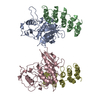 PDB-1bi8: |
| Source |
|
 Keywords Keywords | MEMBRANE PROTEIN / GPCR / cryo-EM / DP1 / BW245C / prostaglandin D2 receptor / prostanoid DP receptor / PGD receptor / PTGDR / Gs protein / DP1-selective agonist / PGD2 / MEMBRANE PROTEIN/Immune System / inverse agonist / ONO2550289 / DP1-bRIL chimera / MEMBRANE PROTEIN-Immune System complex / Apo form / ONO3030297 |


 homo sapiens (human)
