 Movie
Movie Controller
 Structure viewers
 About Yorodumi Papers


+Search query
-Structure paper
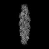
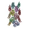
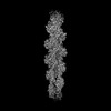
| Title | Distinct inter-domain interactions of dimeric versus monomeric α-catenin link cell junctions to filaments. |
|---|---|
| Journal, issue, pages | Commun Biol, Vol. 6, Issue 1, Page 276, Year 2023 |
| Publish date | Mar 16, 2023 |
 Authors Authors | Erumbi S Rangarajan / Emmanuel W Smith / Tina Izard /  |
| PubMed Abstract | Attachment between cells is crucial for almost all aspects of the life of cells. These inter-cell adhesions are mediated by the binding of transmembrane cadherin receptors of one cell to cadherins of ...Attachment between cells is crucial for almost all aspects of the life of cells. These inter-cell adhesions are mediated by the binding of transmembrane cadherin receptors of one cell to cadherins of a neighboring cell. Inside the cell, cadherin binds β-catenin, which interacts with α-catenin. The transitioning of cells between migration and adhesion is modulated by α-catenin, which links cell junctions and the plasma membrane to the actin cytoskeleton. At cell junctions, a single β-catenin/α-catenin heterodimer slips along filamentous actin in the direction of cytoskeletal tension which unfolds clustered heterodimers to form catch bonds with F-actin. Outside cell junctions, α-catenin dimerizes and links the plasma membrane to F-actin. Under cytoskeletal tension, α-catenin unfolds and forms an asymmetric catch bond with F-actin. To understand the mechanism of this important α-catenin function, we determined the 2.7 Å cryogenic electron microscopy (cryoEM) structures of filamentous actin alone and bound to human dimeric α-catenin. Our structures provide mechanistic insights into the role of the α-catenin interdomain interactions in directing α-catenin function and suggest a bivalent mechanism. Further, our cryoEM structure of human monomeric α-catenin provides mechanistic insights into α-catenin autoinhibition. Collectively, our structures capture the initial α-catenin interaction with F-actin before the sensing of force, which is a crucial event in cell adhesion and human disease. |
 External links External links | Commun Biol / PubMed:36928388 / PubMed Central |
| Methods | EM (helical sym.) / EM (single particle) |
| Resolution | 2.7 - 6.9 Å |
| Structure data | EMDB-26772, PDB-7utj: EMDB-26860, PDB-7uxf:  EMDB-27717: Cryogenic electron microscopy 3D map of human full-length monomeric alpha-catenin |
| Chemicals |  ChemComp-ADP:  ChemComp-MG:  ChemComp-HOH: |
| Source |
|
 Keywords Keywords | CELL ADHESION / alpha-catenin / F-actin / F-actin binding protein / cell-cell junction |

 homo sapiens (human)
