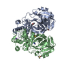
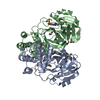
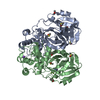
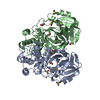
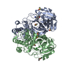
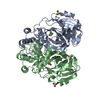
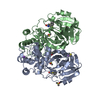
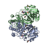
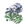
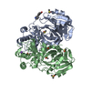
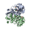
Title Fragment-based screening targeting an open form of the SARS-CoV-2 main protease binding pocket. Journal, issue, pages Acta Crystallogr D Struct Biol , Vol. 80, Page 123-136, Year 2024Publish date Nov 14, 2023 (structure data deposition date) Huang, C.Y. / Metz, A. / Lange, R. / Artico, N. / Potot, C. / Hazemann, J. / Muller, M. / Dos Santos, M. / Chambovey, A. / Ritz, D. ...Huang, C.Y. / Metz, A. / Lange, R. / Artico, N. / Potot, C. / Hazemann, J. / Muller, M. / Dos Santos, M. / Chambovey, A. / Ritz, D. / Eris, D. / Meyer, S. / Bourquin, G. / Sharpe, M. / Mac Sweeney, A. / Methods X-ray diffraction Resolution 1.47 - 1.92 Å Structure data PDB-7gre cpd-1 Method : X-RAY DIFFRACTION / Resolution : 1.66 Å
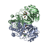
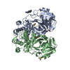
PDB-7grf cpd-2 Method : X-RAY DIFFRACTION / Resolution : 1.84 Å
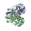
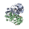
PDB-7grg cpd-3 Method : X-RAY DIFFRACTION / Resolution : 1.54 Å
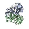
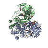
PDB-7grh cpd-4 Method : X-RAY DIFFRACTION / Resolution : 1.87 Å
PDB-7gri cpd-5 Method : X-RAY DIFFRACTION / Resolution : 1.79 Å
PDB-7grj cpd-6 Method : X-RAY DIFFRACTION / Resolution : 1.74 Å
PDB-7grk cpd-7 Method : X-RAY DIFFRACTION / Resolution : 1.8 Å
PDB-7grl cpd-8 Method : X-RAY DIFFRACTION / Resolution : 1.68 Å
PDB-7grm cpd-9 Method : X-RAY DIFFRACTION / Resolution : 1.7 Å
PDB-7grn cpd-10 Method : X-RAY DIFFRACTION / Resolution : 1.92 Å
PDB-7gro cpd-11 Method : X-RAY DIFFRACTION / Resolution : 1.55 Å
PDB-7grp cpd-12 Method : X-RAY DIFFRACTION / Resolution : 1.56 Å
PDB-7grq cpd-13 Method : X-RAY DIFFRACTION / Resolution : 1.67 Å
PDB-7grr cpd-14 Method : X-RAY DIFFRACTION / Resolution : 1.68 Å
PDB-7grs cpd-15 Method : X-RAY DIFFRACTION / Resolution : 1.47 Å
PDB-7grt cpd-16 Method : X-RAY DIFFRACTION / Resolution : 1.71 Å
PDB-7gru cpd-17 Method : X-RAY DIFFRACTION / Resolution : 1.81 Å
PDB-7grv cpd-18 Method : X-RAY DIFFRACTION / Resolution : 1.92 Å
PDB-7grw cpd-19 Method : X-RAY DIFFRACTION / Resolution : 1.92 Å
PDB-7grx cpd-20 Method : X-RAY DIFFRACTION / Resolution : 1.55 Å
PDB-7gry cpd-21 Method : X-RAY DIFFRACTION / Resolution : 1.54 Å
PDB-7grz cpd-22 Method : X-RAY DIFFRACTION / Resolution : 1.86 Å
PDB-7gs0 cpd-23 Method : X-RAY DIFFRACTION / Resolution : 1.69 Å
PDB-7gs1 cpd-24 Method : X-RAY DIFFRACTION / Resolution : 1.74 Å
PDB-7gs2 cpd-25 Method : X-RAY DIFFRACTION / Resolution : 1.77 Å
PDB-7gs3 cpd-26 Method : X-RAY DIFFRACTION / Resolution : 1.89 Å
PDB-7gs4 cpd-27 Method : X-RAY DIFFRACTION / Resolution : 1.86 Å
PDB-7gs5 cpd-28 Method : X-RAY DIFFRACTION / Resolution : 1.88 Å
PDB-7gs6 cpd-29 Method : X-RAY DIFFRACTION / Resolution : 1.62 Å
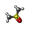
Chemicals ChemComp-DMS / DMSO, precipitant*YM
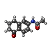
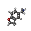
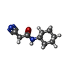
ChemComp-ZHA
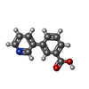
ChemComp-VXQ
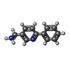
ChemComp-NT9
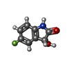
ChemComp-GT7
ChemComp-JAH
ChemComp-0TI
Source / /
 Authors
 External links
 Acta Crystallogr D Struct Biol /
  PubMed:38289714









 Keywords
 Movie
Movie Controller
 Structure viewers
 About Yorodumi Papers


