PDB-5roc: PanDDA analysis group deposition -- Proteinase K crystal structure Apo65 Method: X-RAY DIFFRACTION / Resolution: 1.523 Å
PDB-5rod: PanDDA analysis group deposition -- Proteinase K crystal structure Apo71 Method: X-RAY DIFFRACTION / Resolution: 1.04 Å
PDB-5roe: PanDDA analysis group deposition -- Proteinase K crystal structure Apo6 Method: X-RAY DIFFRACTION / Resolution: 1.5 Å
PDB-5rof: PanDDA analysis group deposition -- Proteinase K changed state model for fragment Frag Xtal Screen C11a Method: X-RAY DIFFRACTION / Resolution: 1.08 Å
PDB-5rog: PanDDA analysis group deposition -- Proteinase K crystal structure Apo51 Method: X-RAY DIFFRACTION / Resolution: 1.08 Å
PDB-5roh: PanDDA analysis group deposition -- Proteinase K crystal structure Apo61 Method: X-RAY DIFFRACTION / Resolution: 1.04 Å
PDB-5roi: PanDDA analysis group deposition -- Proteinase K crystal structure Apo30 Method: X-RAY DIFFRACTION / Resolution: 1.05 Å
PDB-5roj: PanDDA analysis group deposition -- Proteinase K crystal structure Apo59 Method: X-RAY DIFFRACTION / Resolution: 1.02 Å
PDB-5rok: PanDDA analysis group deposition -- Proteinase K crystal structure Apo37 Method: X-RAY DIFFRACTION / Resolution: 1.04 Å
PDB-5rol: PanDDA analysis group deposition -- Proteinase K changed state model for fragment Frag Xtal Screen B5a Method: X-RAY DIFFRACTION / Resolution: 1.22 Å
PDB-5rom: PanDDA analysis group deposition -- Proteinase K crystal structure Apo45 Method: X-RAY DIFFRACTION / Resolution: 1.07 Å
PDB-5ron: PanDDA analysis group deposition -- Proteinase K changed state model for fragment Frag Xtal Screen E4a Method: X-RAY DIFFRACTION / Resolution: 1.22 Å
PDB-5roo: PanDDA analysis group deposition -- Proteinase K crystal structure Apo73 Method: X-RAY DIFFRACTION / Resolution: 1.41 Å
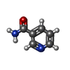
PDB-5rop: PanDDA analysis group deposition -- Proteinase K changed state model for fragment Frag Xtal Screen A12a at Room Temperature Method: X-RAY DIFFRACTION / Resolution: 1.64 Å
PDB-5roq: PanDDA analysis group deposition -- Proteinase K changed state model for fragment Frag Xtal Screen A12a Method: X-RAY DIFFRACTION / Resolution: 1.02 Å
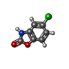
PDB-5ror: PanDDA analysis group deposition -- Proteinase K changed state model for fragment Frag Xtal Screen F1a Method: X-RAY DIFFRACTION / Resolution: 1.22 Å
PDB-5ros: PanDDA analysis group deposition -- Proteinase K crystal structure Apo34 Method: X-RAY DIFFRACTION / Resolution: 1.45 Å
PDB-5rot: PanDDA analysis group deposition -- Proteinase K crystal structure Apo1 Method: X-RAY DIFFRACTION / Resolution: 1.05 Å
PDB-5rou: PanDDA analysis group deposition -- Proteinase K crystal structure Apo72 Method: X-RAY DIFFRACTION / Resolution: 1.06 Å
PDB-5rov: PanDDA analysis group deposition -- Proteinase K crystal structure Apo62 Method: X-RAY DIFFRACTION / Resolution: 1.04 Å
PDB-5row: PanDDA analysis group deposition -- Proteinase K changed state model for fragment Frag Xtal Screen F6a Method: X-RAY DIFFRACTION / Resolution: 1.21 Å
PDB-5rox: PanDDA analysis group deposition -- Proteinase K crystal structure Apo24 Method: X-RAY DIFFRACTION / Resolution: 1.12 Å
PDB-5roy: PanDDA analysis group deposition -- Proteinase K crystal structure Apo22 Method: X-RAY DIFFRACTION / Resolution: 1.05 Å
PDB-5roz: PanDDA analysis group deposition -- Proteinase K crystal structure Apo41 Method: X-RAY DIFFRACTION / Resolution: 1.14 Å
PDB-5rp0: PanDDA analysis group deposition -- Proteinase K crystal structure Apo57 Method: X-RAY DIFFRACTION / Resolution: 1.03 Å
PDB-5rp1: PanDDA analysis group deposition -- Proteinase K crystal structure Apo36 Method: X-RAY DIFFRACTION / Resolution: 1.11 Å
PDB-5rp2: PanDDA analysis group deposition -- Proteinase K crystal structure Apo20 Method: X-RAY DIFFRACTION / Resolution: 1.1 Å
PDB-5rp3: PanDDA analysis group deposition -- Proteinase K crystal structure Apo14 Method: X-RAY DIFFRACTION / Resolution: 1.09 Å
PDB-5rp4: PanDDA analysis group deposition -- Proteinase K crystal structure Apo70 Method: X-RAY DIFFRACTION / Resolution: 1.05 Å
PDB-5rp5: PanDDA analysis group deposition -- Proteinase K crystal structure Apo48 Method: X-RAY DIFFRACTION / Resolution: 1.09 Å
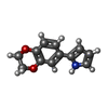
PDB-5rp6: PanDDA analysis group deposition -- Proteinase K changed state model for fragment Frag Xtal Screen C2a Method: X-RAY DIFFRACTION / Resolution: 1.17 Å
PDB-5rp7: PanDDA analysis group deposition -- Proteinase K changed state model for fragment Frag Xtal Screen D12a Method: X-RAY DIFFRACTION / Resolution: 1.15 Å
PDB-5rp8: PanDDA analysis group deposition -- Proteinase K crystal structure Apo46 Method: X-RAY DIFFRACTION / Resolution: 1.03 Å
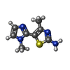
PDB-5rp9: PanDDA analysis group deposition -- Proteinase K changed state model for fragment Frag Xtal Screen B7a Method: X-RAY DIFFRACTION / Resolution: 1.48 Å
PDB-5rpa: PanDDA analysis group deposition -- Proteinase K changed state model for fragment Frag Xtal Screen E3a Method: X-RAY DIFFRACTION / Resolution: 1.25 Å
PDB-5rpb: PanDDA analysis group deposition -- Proteinase K crystal structure Apo68 Method: X-RAY DIFFRACTION / Resolution: 1.64 Å
PDB-5rpc: PanDDA analysis group deposition -- Proteinase K changed state model for fragment Frag Xtal Screen H3a Method: X-RAY DIFFRACTION / Resolution: 1.23 Å
PDB-5rpd: PanDDA analysis group deposition -- Proteinase K changed state model for fragment Frag Xtal Screen F12a Method: X-RAY DIFFRACTION / Resolution: 1.02 Å
PDB-5rpe: PanDDA analysis group deposition -- Proteinase K crystal structure Apo40 Method: X-RAY DIFFRACTION / Resolution: 1.02 Å
PDB-5rpf: PanDDA analysis group deposition -- Proteinase K crystal structure Apo27 Method: X-RAY DIFFRACTION / Resolution: 1.39 Å
PDB-5rpg: PanDDA analysis group deposition -- Proteinase K changed state model for fragment Frag Xtal Screen H2a Method: X-RAY DIFFRACTION / Resolution: 1.5 Å
PDB-5rph: PanDDA analysis group deposition -- Proteinase K changed state model for fragment Frag Xtal Screen A11a Method: X-RAY DIFFRACTION / Resolution: 1.38 Å
PDB-5rpi: PanDDA analysis group deposition -- Proteinase K crystal structure Apo26 Method: X-RAY DIFFRACTION / Resolution: 1.03 Å
PDB-5rpj: PanDDA analysis group deposition -- Proteinase K changed state model for fragment Frag Xtal Screen B12a Method: X-RAY DIFFRACTION / Resolution: 1.27 Å
PDB-5rpk: PanDDA analysis group deposition -- Proteinase K changed state model for fragment Frag Xtal Screen B9a Method: X-RAY DIFFRACTION / Resolution: 1.78 Å
PDB-5rpl: PanDDA analysis group deposition -- Proteinase K changed state model for fragment Frag Xtal Screen C8a Method: X-RAY DIFFRACTION / Resolution: 1.09 Å
PDB-5rpm: PanDDA analysis group deposition -- Proteinase K changed state model for fragment Frag Xtal Screen H5a Method: X-RAY DIFFRACTION / Resolution: 1.22 Å
PDB-5rpn: PanDDA analysis group deposition -- Proteinase K crystal structure Apo64 Method: X-RAY DIFFRACTION / Resolution: 1.02 Å
PDB-5rpo: PanDDA analysis group deposition -- Proteinase K crystal structure Apo7 Method: X-RAY DIFFRACTION / Resolution: 1.5 Å
PDB-5rpp: PanDDA analysis group deposition -- Proteinase K crystal structure Apo44 Method: X-RAY DIFFRACTION / Resolution: 1.08 Å
PDB-5rpq: PanDDA analysis group deposition -- Proteinase K crystal structure Apo32 Method: X-RAY DIFFRACTION / Resolution: 1.07 Å
PDB-5rpr: PanDDA analysis group deposition -- Proteinase K crystal structure Apo15 Method: X-RAY DIFFRACTION / Resolution: 1.16 Å
PDB-5rps: PanDDA analysis group deposition -- Proteinase K crystal structure Apo60 Method: X-RAY DIFFRACTION / Resolution: 1.04 Å
PDB-5rpt: PanDDA analysis group deposition -- Proteinase K crystal structure Apo9 Method: X-RAY DIFFRACTION / Resolution: 1.98 Å
PDB-5rpu: PanDDA analysis group deposition -- Proteinase K crystal structure Apo67 Method: X-RAY DIFFRACTION / Resolution: 1.55 Å
PDB-5rpv: PanDDA analysis group deposition -- Proteinase K crystal structure Apo35 Method: X-RAY DIFFRACTION / Resolution: 1.1 Å
PDB-5rpw: PanDDA analysis group deposition -- Proteinase K crystal structure Apo63 Method: X-RAY DIFFRACTION / Resolution: 1.02 Å
PDB-5rpx: PanDDA analysis group deposition -- Proteinase K crystal structure Apo43 Method: X-RAY DIFFRACTION / Resolution: 1.05 Å
PDB-5rpy: PanDDA analysis group deposition -- Proteinase K crystal structure Apo10 Method: X-RAY DIFFRACTION / Resolution: 1.09 Å
PDB-5rpz: PanDDA analysis group deposition -- Proteinase K crystal structure Apo58 Method: X-RAY DIFFRACTION / Resolution: 1.22 Å
In the structure databanks used in Yorodumi, some data are registered as the other names, "COVID-19 virus" and "2019-nCoV". Here are the details of the virus and the list of structure data.
Jan 31, 2019. EMDB accession codes are about to change! (news from PDBe EMDB page)
EMDB accession codes are about to change! (news from PDBe EMDB page)
The allocation of 4 digits for EMDB accession codes will soon come to an end. Whilst these codes will remain in use, new EMDB accession codes will include an additional digit and will expand incrementally as the available range of codes is exhausted. The current 4-digit format prefixed with “EMD-” (i.e. EMD-XXXX) will advance to a 5-digit format (i.e. EMD-XXXXX), and so on. It is currently estimated that the 4-digit codes will be depleted around Spring 2019, at which point the 5-digit format will come into force.
The EM Navigator/Yorodumi systems omit the EMD- prefix.
Related info.:Q: What is EMD? / ID/Accession-code notation in Yorodumi/EM Navigator
 Movie
Movie Controller
 Structure viewers
 About Yorodumi Papers


 Authors
 External links


 Keywords
 parengyodontium album (fungus)