 Movie
Movie Controller
 Structure viewers
 About Yorodumi Papers


+Search query
-Structure paper
| Title | Reovirus polymerase lambda 3 localized by cryo-electron microscopy of virions at a resolution of 7.6 A. |
|---|---|
| Journal, issue, pages | Nat Struct Biol, Vol. 10, Issue 12, Page 1011-1018, Year 2003 |
| Publish date | Nov 9, 2003 |
 Authors Authors | Xing Zhang / Stephen B Walker / Paul R Chipman / Max L Nibert / Timothy S Baker /  |
| PubMed Abstract | Reovirus is an icosahedral, double-stranded (ds) RNA virus that uses viral polymerases packaged within the viral core to transcribe its ten distinct plus-strand RNAs. To localize these polymerases, ...Reovirus is an icosahedral, double-stranded (ds) RNA virus that uses viral polymerases packaged within the viral core to transcribe its ten distinct plus-strand RNAs. To localize these polymerases, the structure of the reovirion was refined to a resolution of 7.6 A by cryo-electron microscopy (cryo-EM) and three-dimensional (3D) image reconstruction. X-ray crystal models of reovirus proteins, including polymerase lambda 3, were then fitted into the density map. Each copy of lambda 3 was found anchored to the inner surface of the icosahedral core shell, making major contacts with three molecules of shell protein lambda 1 and overlapping, but not centering on, a five-fold axis. The overlap explains why only one copy of lambda 3 is bound per vertex. lambda 3 is furthermore oriented with its transcript exit channel facing a small channel through the lambda 1 shell, suggesting how the nascent RNA is passed into the large external cavity of the pentameric capping enzyme complex formed by protein lambda 2. |
 External links External links | Nat Struct Biol / PubMed:14608373 / PubMed Central |
| Methods | EM (single particle) |
| Resolution | 7.6 Å |
| Structure data | 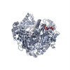 PDB-1uon: |
| Chemicals |  ChemComp-CH1:  ChemComp-MN:  ChemComp-HOH: |
| Source |
|
 Keywords Keywords | POLYMERASE / REOVIRUS / CRYOEM / CORE PROTEIN |
 reovirus