 Movie
Movie Controller
 Structure viewers
 About Yorodumi Papers


+Search query
-Structure paper
| Title | Model for the structure of bacteriorhodopsin based on high-resolution electron cryo-microscopy. |
|---|---|
| Journal, issue, pages | J Mol Biol, Vol. 213, Issue 4, Page 899-929, Year 1990 |
| Publish date | Jun 20, 1990 |
 Authors Authors | R Henderson / J M Baldwin / T A Ceska / F Zemlin / E Beckmann / K H Downing /  |
| PubMed Abstract | The light-driven proton pump bacteriorhodopsin occurs naturally as two-dimensional crystals. A three-dimensional density map of the structure, at near-atomic resolution, has been obtained by studying ...The light-driven proton pump bacteriorhodopsin occurs naturally as two-dimensional crystals. A three-dimensional density map of the structure, at near-atomic resolution, has been obtained by studying the crystals using electron cryo-microscopy to obtain electron diffraction patterns and high-resolution micrographs. New methods were developed for analysing micrographs from tilted specimens, incorporating methods previously developed for untilted specimens that enable large areas to be analysed and corrected for distortions. Data from 72 images, from both tilted and untilted specimens, were analysed to produce the phases of 2700 independent Fourier components of the structure. The amplitudes of these components were accurately measured from 150 diffraction patterns. Together, these data represent about half of the full three-dimensional transform to 3.5 A. The map of the structure has a resolution of 3.5 A in a direction parallel to the membrane plane but lower than this in the perpendicular direction. It shows many features in the density that are resolved from the main density of the seven alpha-helices. We interpret these features as the bulky aromatic side-chains of phenylalanine, tyrosine and tryptophan residues. There is also a very dense feature, which is the beta-ionone ring of the retinal chromophore. Using these bulky side-chains as guide points and taking account of bulges in the helices that indicate smaller side-chains such as leucine, a complete atomic model for bacteriorhodopsin between amino acid residues 8 and 225 has been built. There are 21 amino acid residues, contributed by all seven helices, surrounding the retinal and 26 residues, contributed by five helices, forming the proton pathway or channel. Ten of the amino acid residues in the middle of the proton channel are also part of the retinal binding site. The model also provides a useful basis for consideration of the mechanism of proton pumping and allows a consistent interpretation of a great deal of other experimental data. In particular, the structure suggests that pK changes in the Schiff base must act as the means by which light energy is converted into proton pumping pressure in the channel. Asp96 is on the pathway from the cytoplasm to the Schiff base and Asp85 is on the pathway from the Schiff base to the extracellular surface. |
 External links External links | J Mol Biol / PubMed:2359127 |
| Methods | EM (electron crystallography) |
| Resolution | 3.5 Å |
| Structure data | 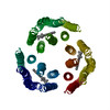 PDB-1brd: |
| Chemicals |  ChemComp-RET: |
| Source |
|
 Keywords Keywords | PHOTORECEPTOR |
 halobacterium salinarum (Halophile)