[English] 日本語
Yorodumi Papers
- Database of articles cited by EMDB/PDB/SASBDB data -

+
Search query

Keywords
Structure methods
Author
Journal
IF

-
Structure paper

TitleActivation mechanism and structural assembly of the Mycobacterium tuberculosis ClpP1P2 protease and its associated ATPases.
Journal, issue, pagesCell Rep, Vol. 45, Issue 6, Page 117400, Year 2026
Publish dateJun 23, 2026
AuthorsKatharina Weinhäupl / Tatos Akopian / Dmitry A Semchonok / Olga Krandor / Rocío Arranz / M Teresa Bueno-Carrasco / Marcos Gragera / Maelenn Chevreuil / Bertrand Raynal / Samuel Zinga / Yuxin Liu / Jack Lai / WenGen Wu / William Bachovchin / Diego Gauto / Alfred Goldberg / Eric Rubin / Hugo Fraga /
PubMed AbstractSupramolecular assemblies are fundamental to cellular biochemical processes, relying on their dynamic nature to perform essential functions. The protease ClpP1P2, in association with ATPase partners ...Supramolecular assemblies are fundamental to cellular biochemical processes, relying on their dynamic nature to perform essential functions. The protease ClpP1P2, in association with ATPase partners ClpC1 or ClpX, is critical for the survival of Mycobacterium tuberculosis (Mtb). While the ClpP1P2 complex requires activation by specific N-blocked dipeptides to exhibit proteolytic activity in vitro, the mechanism of in vivo activation remains unclear. In this study, we use cryo-electron microscopy (cryo-EM) to determine the structure of the ClpC1P1P2 complex, revealing a highly asymmetric architecture with ClpC1 bound to the ClpP1P2 protease barrel. The activator dipeptide is observed only in the ClpP2 active site, while the ClpP1 entry pore remains closed. Molecular crowding agents promote the formation of larger ClpXP1P2 and ClpC1P1P2 complexes, enhancing structural stability and enzymatic activity. These findings suggest that molecular crowding stabilizes these complexes and promotes their activation, providing new insights into ClpC1P1P2 structural dynamics and function.
External linksCell Rep / PubMed:42213777
MethodsEM (single particle)
Resolution3.05 - 3.37 Å
Structure data

EMDB-52764: Structure of the Mycobacterium tuberculosis ClpC1P1P2 complex bound to the activator Bz-LL - focused refinement ClpC1
Method: EM (single particle) / Resolution: 3.13 Å

EMDB-52765: Structure of the Mycobacterium tuberculosis ClpC1P1P2 complex bound to the activator Bz-LL - consensus map
Method: EM (single particle) / Resolution: 3.37 Å

EMDB-52766: Structure of the Mycobacterium tuberculosis ClpC1P1P2 complex bound to the activator Bz-LL - focused map ClpP1P2
Method: EM (single particle) / Resolution: 3.05 Å

EMDB-52840, PDB-9if4:
Structure of the Mycobacterium Tuberculosis ClpC1P1P2 complex bound to the activator Bz-Leu-Leu
Method: EM (single particle) / Resolution: 3.09 Å

Chemicals

ChemComp-ADP:
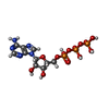
ADENOSINE-5'-DIPHOSPHATE / ADP, energy-carrying molecule*YM

ChemComp-ATP:
ADENOSINE-5'-TRIPHOSPHATE / ATP, energy-carrying molecule*YM

Source
  • mycobacterium tuberculosis (bacteria)
  • synthetic construct (others)
KeywordsCHAPERONE / protein quality control / peptide activator / protease / ATPase

+
About Yorodumi Papers

-
News

-
Feb 9, 2022. New format data for meta-information of EMDB entries

New format data for meta-information of EMDB entries

  • Version 3 of the EMDB header file is now the official format.
  • The previous official version 1.9 will be removed from the archive.

Related info.:EMDB header

External links:wwPDB to switch to version 3 of the EMDB data model

-
Aug 12, 2020. Covid-19 info

Covid-19 info

URL: https://pdbj.org/emnavi/covid19.php

New page: Covid-19 featured information page in EM Navigator.

Related info.:Covid-19 info / Mar 5, 2020. Novel coronavirus structure data

+
Mar 5, 2020. Novel coronavirus structure data

Novel coronavirus structure data

Related info.:Yorodumi Speices / Aug 12, 2020. Covid-19 info

External links:COVID-19 featured content - PDBj / Molecule of the Month (242):Coronavirus Proteases

+
Jan 31, 2019. EMDB accession codes are about to change! (news from PDBe EMDB page)

EMDB accession codes are about to change! (news from PDBe EMDB page)

  • The allocation of 4 digits for EMDB accession codes will soon come to an end. Whilst these codes will remain in use, new EMDB accession codes will include an additional digit and will expand incrementally as the available range of codes is exhausted. The current 4-digit format prefixed with “EMD-” (i.e. EMD-XXXX) will advance to a 5-digit format (i.e. EMD-XXXXX), and so on. It is currently estimated that the 4-digit codes will be depleted around Spring 2019, at which point the 5-digit format will come into force.
  • The EM Navigator/Yorodumi systems omit the EMD- prefix.

Related info.:Q: What is EMD? / ID/Accession-code notation in Yorodumi/EM Navigator

External links:EMDB Accession Codes are Changing Soon! / Contact to PDBj

+
Jul 12, 2017. Major update of PDB

Major update of PDB

  • wwPDB released updated PDB data conforming to the new PDBx/mmCIF dictionary.
  • This is a major update changing the version number from 4 to 5, and with Remediation, in which all the entries are updated.
  • In this update, many items about electron microscopy experimental information are reorganized (e.g. em_software).
  • Now, EM Navigator and Yorodumi are based on the updated data.

External links:wwPDB Remediation / Enriched Model Files Conforming to OneDep Data Standards Now Available in the PDB FTP Archive

-
Yorodumi Papers

Database of articles cited by EMDB/PDB/SASBDB data

  • Database of articles cited by EMDB, PDB, and SASBDB entries
  • Using PubMed data

Related info.:EMDB / PDB / SASBDB / Yorodumi / EMN Papers / Changes in new EM Navigator and Yorodumi

Read more