 Movie
Movie Controller
 Structure viewers
 About Yorodumi Papers


+Search query
-Structure paper
| Title | Reducing the effects of radiation damage in cryo-EM using liquid helium temperatures. |
|---|---|
| Journal, issue, pages | Proc Natl Acad Sci U S A, Vol. 122, Issue 17, Page e2421538122, Year 2025 |
| Publish date | Apr 29, 2025 |
 Authors Authors | Joshua L Dickerson / Katerina Naydenova / Mathew J Peet / Hugh Wilson / Biplob Nandy / Greg McMullan / Robert Morrison / Christopher J Russo /  |
| PubMed Abstract | The physical limit in determining the atomic structure of biological molecules is radiation damage. In electron cryomicroscopy, there have been numerous attempts to reduce the effects of radiation ...The physical limit in determining the atomic structure of biological molecules is radiation damage. In electron cryomicroscopy, there have been numerous attempts to reduce the effects of radiation damage by cooling the specimen beyond liquid-nitrogen temperatures, yet all failed to realize the potential improvement for single-particle structure determination. We have identified the physical causes of information loss at liquid-helium temperatures, and overcome them using a combination of nanoscale electron beam illumination and a gold specimen support with 100 nm diameter holes. This combination allowed structure determination where every frame in the exposure contained more information than was available with cryomicroscopy at liquid-nitrogen temperatures, matching expectations from crystal diffraction. Since a 100 nm hole is smaller than the field of view of a typical micrograph, the edges of the foil are directly visible in each micrograph. Protein molecules that are degraded tend to aggregate at the edges of foil holes and can constitute a significant fraction of the micrograph. This and the need for minimal water-foil irradiation will both be important to consider as new cryomicroscopes and specimen supports are developed for imaging molecules at extremely low temperatures where the effects of radiation damage are reduced. |
 External links External links | Proc Natl Acad Sci U S A / PubMed:40261934 / PubMed Central |
| Methods | EM (single particle) |
| Resolution | 2.1 - 3.32 Å |
| Structure data | 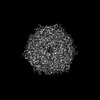 EMDB-51777: Structure of DPS determined at 13K in 100 nm diameter holes. 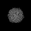 EMDB-51778: Structure of DPS determined at 81K in 100 nm diameter holes. 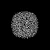 EMDB-51779: Structure of apoferritin determined at 13K in 100 nm diameter holes. 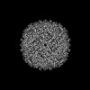 EMDB-51780: Structure of apoferritin determined at 81K in 100 nm diameter holes. 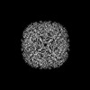 EMDB-51781: Structure of apoferritin determined at 13K in 100 nm diameter holes. 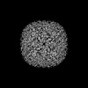 EMDB-51782: Structure of apoferritin determined at 81K in 100 nm diameter holes. 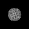 EMDB-51783: Structure of apoferritin determined at 13K in 300 nm diameter holes. 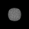 EMDB-51784: Structure of apoferritin determined at 81K in 300 nm diameter holes. |
| Source |
|


